










Giant Cell Arteritis (Temporal Arteritis): Symptoms, Tests and Treatment
The essentials
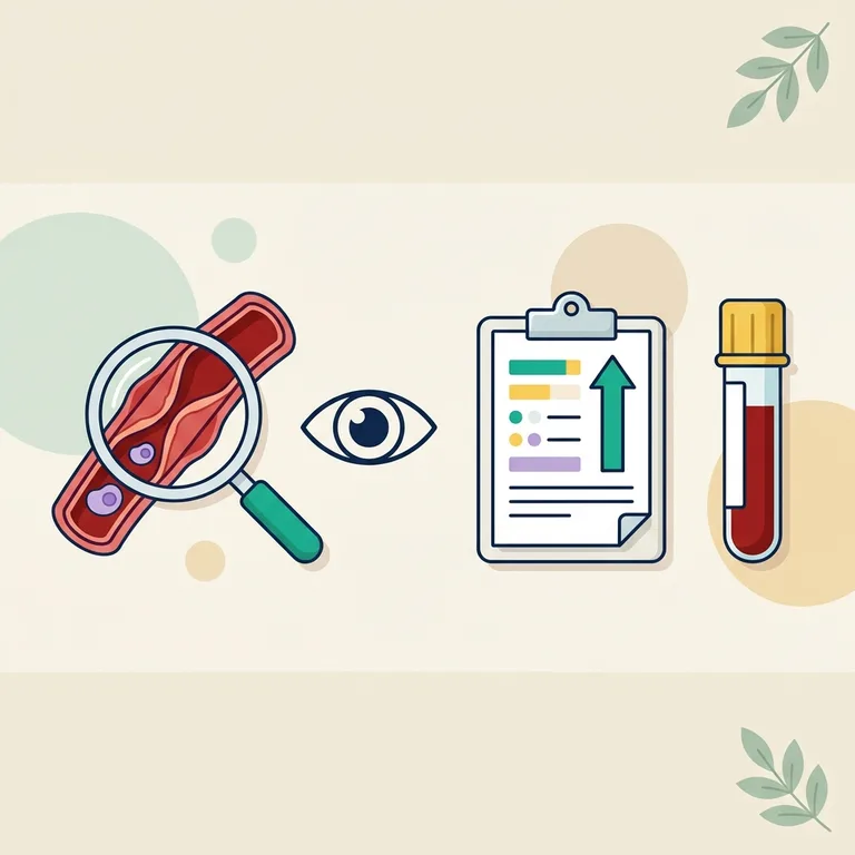
Giant cell arteritis, also called temporal arteritis or Horton disease, is an inflammation of large arteries that only affects people over 50. It typically causes a new headache at the temples, scalp tenderness and jaw pain when chewing. The sed rate and CRP are almost always high, but only an ultrasound, a biopsy or another scan can confirm it. Any change in vision is an emergency: quick treatment can save your sight.
What is giant cell arteritis, at a glance?
Giant cell arteritis (GCA) is a vasculitis: the immune system inflames the wall of large and medium arteries, mainly those supplying the scalp, the jaw and the eyes, and sometimes the aorta. The swollen wall narrows the artery and can cut off blood flow[5].
| Topic | Key facts |
|---|---|
| Other names | Temporal arteritis, Horton disease (the usual name in French-speaking countries)[5] |
| Who gets it | Only adults aged 50 or more, usually around 70; women three times as often as men; mostly people of European descent[5] |
| How common | About 10 new cases per 100,000 people over 50 each year, 11 in the Americas and 22 in Scandinavia[7] |
| Key tests | Sed rate (ESR), CRP, blood count, ultrasound of the temporal arteries, temporal artery biopsy, sometimes a PET scan |
What are the symptoms of giant cell arteritis?
- Common A new headache, often at the temples, unlike any headache you have had before[10].
- Common Scalp tenderness when brushing your hair or lying on a pillow; the temple artery may be thick or tender[5].
- Common Fatigue, weight loss, low fever, sometimes as the only signs[10].
- Possible Jaw claudication: jaw pain that comes on while chewing and eases at rest[5].
- Possible Shoulder and hip pain with morning stiffness: polymyalgia rheumatica (PMR), present in 30 to 50% of people with GCA[5].
- Possible Vision problems: blurred or double vision, a curtain over one eye, sudden loss of sight. In about 30% of cases, brief warning episodes come first[5].
- Rare Stroke, in 2.7 to 7.4% of patients, mostly early on[5].
Most headaches are not GCA: a migraine starts much earlier in life, and face pain can come from a nerve, as in trigeminal neuralgia.
When should I see a doctor?
When to see a doctor
- Emergency: call 911 or go to the emergency room for any sudden loss of vision, double vision or dark curtain over one eye, even if it lasted only minutes, or for signs of stroke. Lost sight may not come back, and the other eye can follow quickly[5].
- The same day if you are over 50 with a new headache, scalp tenderness or jaw pain when chewing. European guidelines ask for a specialist assessment within 24 hours[1].
- At your next appointment for shoulder and hip stiffness, or a high sed rate with tiredness but no headache or eye symptoms.
What causes giant cell arteritis, and who is at risk?
The cause is unknown. Immune cells invade the artery wall and release inflammatory messengers, especially interleukin-6, which thicken the wall[5]. Risk rises with age (it never starts before 50 and peaks after 70), female sex, and Northern European origin: the disease is 2 to 5 times more frequent in Northern than in Southern Europe and much rarer in people of African, Asian or Arab origin[5]. It belongs to the family described in our guide to autoimmune diseases and their blood tests.
How is giant cell arteritis diagnosed, and what do the blood tests show?
No blood test proves GCA on its own[10]. Doctors combine, after 50, typical symptoms, blood inflammation (in more than 95% of cases) and proof of vasculitis on imaging or biopsy[5].
- CRP and sed rate (ESR): in 764 biopsied patients, CRP was high in 87% of those with GCA and ESR in 84%. Yet 4% had both normal[6]: a normal result never cancels an eye emergency.
- Complete blood count: about half of patients have mild anemia and a high platelet count, both caused by inflammation[5].
- Liver tests: a raised alkaline phosphatase appears in about one patient in three[5].
- Fibrinogen: France prefers it to the ESR, which anemia alone can raise[5].
| Test | Result | Reference range | Flag |
|---|---|---|---|
| Sed rate (ESR), Westergren | 78 mm/hr | 0-30 mm/hr | H |
| C-reactive protein (CRP) | 6.8 mg/dL | < 1.0 mg/dL | H |
| Hemoglobin | 11.2 g/dL | 12.0-15.5 g/dL | L |
| Platelet count | 498 x103/µL | 150-450 x103/µL | H |
| Alkaline phosphatase (ALP) | 162 U/L | 44-147 U/L | H |
- ESR 78 mm/hr: for women aged 50 and over, the usual limit is 30 mm/hr; very high values occur with GCA, but also with infections and some cancers[11].
- CRP 6.8 mg/dL (68 mg/L): healthy CRP is about 1.0 mg/dL or lower. CRP shows how much inflammation there is, not where it comes from[12].
- Hemoglobin L, platelets H, ALP H: the pattern of inflammation often seen in GCA[5], though an infection could give the same report. Your own lab's ranges apply.
A very high sed rate and a headache that worries you? BloodSense explains every line of your report, ESR, CRP, platelets and more, so you know what to ask and how urgently.
Get my blood results explainedUltrasound and the "halo sign"
Since 2023, European guidelines recommend ultrasound of the temporal and armpit (axillary) arteries as the first imaging test for suspected GCA[2]. The typical finding is the halo sign: a dark, even ring of swollen wall around the artery. Its reliability depends on the operator's experience[5].
Biopsy, PET scan and MRI
A temporal artery biopsy, done under local anesthesia, is the only test that shows the vasculitis under the microscope[5]. The 2021 American guideline prefers it to ultrasound, with a segment longer than 1 cm[3]. A PET scan, MRI or CT angiogram can show inflammation of the aorta, present in about 40% of patients[5], and helps when the biopsy is negative[2][3].
For research, the 2022 ACR/EULAR score gives 5 points for a positive biopsy or halo sign and 3 points for an ESR of 50 mm/hr or more or a CRP of 10 mg/L or more; 6 points classify GCA[4]. It is a research tool, not a diagnosis.
Which tests are used to follow giant cell arteritis under treatment?
Under steroids, CRP usually returns to normal within 1 to 3 weeks[5]. Follow-up then checks three things:
- Disease activity: CRP and a slow marker (ESR or fibrinogen), about monthly at first; visits every 1 to 3 months for 3 months, then every 3 to 6 months[5]. Almost half of patients relapse, mostly within 2 years[9]. A rising marker without symptoms usually means closer watching, not automatically more treatment[3].
- Steroid side effects: potassium after 1 to 2 weeks, blood sugar at 1 month, then HbA1c and cholesterol every 3 months[5]; bone protection from the start against osteoporosis[10].
- Tocilizumab safety: blood count and liver enzymes every 1 to 2 weeks the first month, then every 1 to 3 months, and a lipid panel after 8 to 12 weeks[5].
Watch out: tocilizumab blocks interleukin-6, the signal that makes the liver produce CRP. Under this treatment, CRP and ESR usually stay low even during a relapse or an infection[5], so new symptoms count more than a normal result. When the aorta is involved, imaging is repeated over the years to detect an aneurysm, which can appear late[5].
How is giant cell arteritis treated?
With a strong suspicion, treatment starts at once, without waiting for confirmation tests[1]. No doses are given here.
- Corticosteroids (prednisone): the first treatment. Most people improve within days; the dose is lowered slowly over 1 to 2 years[10]. When vision is threatened, intravenous steroids may come first[3].
- Tocilizumab: an injected biologic that blocks the interleukin-6 receptor. In the main trial, 53 to 56% of treated patients were in lasting remission at one year, against 14 to 18% on steroids alone, with about half the prednisone[8]. About half relapse in the year after stopping it[5].
- Other options: the 2025 EULAR update adds upadacitinib, a tablet, for selected patients, with methotrexate as an alternative[1].
- Supportive care: calcium, vitamin D, sometimes a bisphosphonate or aspirin[10].
How can I live well with giant cell arteritis?
- Never stop steroids suddenly or lower the dose on your own.
- Know the signs of a relapse: headache, jaw pain, shoulder stiffness and, above all, any change in vision.
- Report any fever quickly: treatment lowers your defenses and can mask infection[5].
- Control blood pressure and cholesterol and avoid smoking: cardiovascular risk stays higher for years[5]. Know the signs of a stroke.
Questions to ask your doctor
- Should I start steroids now, before the ultrasound or biopsy?
- Do I also have polymyalgia rheumatica?
- Which blood tests will check my treatment, and how often?
- If I take tocilizumab, how will we detect a relapse?
Frequently asked questions
Can giant cell arteritis be cured?
Most people reach lasting remission and can stop treatment, usually after 1 to 2 years or more. It can come back, so follow-up continues[10].
Is vision loss from giant cell arteritis reversible?
Usually not. Treatment mainly protects the other eye, which is why steroids are started without waiting[1][5].
What is the difference with polymyalgia rheumatica?
PMR inflames the shoulders and hips, not the arteries, and needs lower steroid doses. The two often coexist[5].
Can you die from giant cell arteritis?
Deaths are rare with early treatment, and mortality has fallen over the decades[7]. The serious risks are stroke at onset and aortic complications later[5].
Is Horton disease the same as Horton's headache?
No. "Horton's headache" is an old name for cluster headache, a different disorder of younger adults with short, intense attacks around one eye.
Related articles
- Sed rate (ESR)How to read a high sed rate and its causes
- CRP blood testWhat a high inflammation marker means
- Platelet countWhy platelets rise with inflammation
- ANCA blood testThe test for other types of vasculitis
- FibromyalgiaWidespread pain with normal inflammation tests
- Mukhtyar CB, Nikiphorou E, Bartoletti A, et al. 2025 EULAR recommendations for the management of polymyalgia rheumatica and primary large vessel vasculitis. Ann Rheum Dis. 2026 (online July 21, 2026). doi:10.1016/j.ard.2026.06.009 (accessed November 7, 2026)
- Dejaco C, Ramiro S, Bond M, et al. EULAR recommendations for the use of imaging in large vessel vasculitis in clinical practice: 2023 update. Ann Rheum Dis. 2024;83(6):741-751. doi:10.1136/ard-2023-224543 (accessed November 7, 2026)
- Maz M, Chung SA, Abril A, et al. 2021 American College of Rheumatology/Vasculitis Foundation Guideline for the Management of Giant Cell Arteritis and Takayasu Arteritis. Arthritis Rheumatol. 2021;73(8):1349-1365. doi:10.1002/art.41774. PubMed Central (accessed November 7, 2026)
- Ponte C, Grayson PC, Robson JC, et al. 2022 American College of Rheumatology/EULAR classification criteria for giant cell arteritis. Ann Rheum Dis. 2022;81(12):1647-1653. doi:10.1136/ard-2022-223480 (accessed November 7, 2026)
- Haute Autorité de Santé (France), GEFA, FAI²R. Protocole national de diagnostic et de soins (PNDS) : artérite à cellules géantes (Horton). January 2024. has-sante.fr (in French; accessed November 7, 2026)
- Kermani TA, Schmidt J, Crowson CS, et al. Utility of erythrocyte sedimentation rate and C-reactive protein for the diagnosis of giant cell arteritis. Semin Arthritis Rheum. 2012;41(6):866-871. doi:10.1016/j.semarthrit.2011.10.005 (accessed November 7, 2026)
- Li KJ, Semenov D, Turk M, Pope J. A meta-analysis of the epidemiology of giant cell arteritis across time and space. Arthritis Res Ther. 2021;23(1):82. doi:10.1186/s13075-021-02450-w (accessed November 7, 2026)
- Stone JH, Tuckwell K, Dimonaco S, et al. Trial of tocilizumab in giant-cell arteritis. N Engl J Med. 2017;377(4):317-328. doi:10.1056/NEJMoa1613849. PubMed (accessed November 7, 2026)
- Moreel L, Betrains A, Molenberghs G, et al. Epidemiology and predictors of relapse in giant cell arteritis: a systematic review and meta-analysis. Joint Bone Spine. 2023;90(1):105494. doi:10.1016/j.jbspin.2022.105494 (accessed November 7, 2026)
- MedlinePlus Medical Encyclopedia. Giant cell arteritis. US National Library of Medicine. medlineplus.gov (accessed November 7, 2026)
- MedlinePlus Medical Encyclopedia. Sed rate (erythrocyte sedimentation rate). US National Library of Medicine. medlineplus.gov (accessed November 7, 2026)
- MedlinePlus. C-Reactive Protein (CRP) Test. US National Library of Medicine. medlineplus.gov (accessed November 7, 2026)
This page is general information. It does not replace the advice of a doctor; a blood test alone can neither confirm nor rule out giant cell arteritis. Any change in vision needs emergency care.