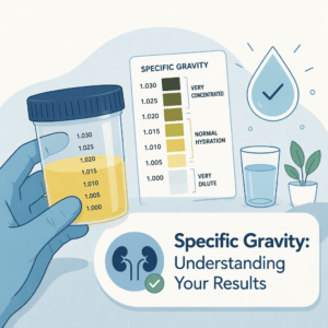
Urine microscopy plays an essential role in assessing kidney and urinary tract health. This test involves examining urine components under a microscope to identify cells, crystals, bacteria, and other substances. As a non-invasive tool, urine microscopy provides valuable clues about infection, inflammation, and kidney function. Understanding what urine microscopy reveals and how it works helps patients appreciate its significance.
What is urine microscopy?
Urine microscopy refers to the microscopic analysis of substances found in urine. Urine itself forms through a filtration process in the kidneys, where blood waste products and excess substances pass into urine. The urine contains water, salts, metabolic waste, cells, and sometimes crystals or microbes. Microscopically, urine can show red and white blood cells, epithelial cells from the urinary tract lining, bacteria, and various crystal types.
Think of urine microscopy as a tiny investigative tool that examines the “ingredients” in urine. It distinguishes normal particles from those that indicate disease. The test usually focuses on three subtypes: cellular elements (like blood or epithelial cells), crystals (which may indicate kidney stones), and microorganisms (bacteria or fungi). Each subtype provides specific diagnostic information.
Behind the scenes: the biology of urine microscopy
Urine forms through complex kidney filtration called glomerular filtration, tubular reabsorption, and secretion. Cells from the urinary tract naturally shed into urine. When damage or infection occurs, more cells—especially white blood cells or red blood cells—enter the urine. Crystals form when substances like calcium or uric acid reach high concentrations and crystallize.
Imagine the urinary system as a water filter and canal. Under normal conditions, only filtered waste and minimal cells pass through. If the filter or canal lining becomes inflamed or injured, extra cells spill into urine. Bacteria often enter from the urethra and multiply, triggering infection signals seen in microscopy.
Biochemically, urine composition shifts according to bodily metabolic activities, hydration status, and pathological processes. For example, urinary tract infections raise white blood cell counts, while kidney stones produce crystals shaped like tiny stones. These microscopic changes reveal ongoing biological events inside the body.
The urine microscopy test: before, during, and after
Doctors order urine microscopy to diagnose urinary tract infections, kidney diseases, or unexplained urinary symptoms such as blood in the urine or pain. Sometimes, the test forms part of routine health screens or hospital admission labs.
Before the test, no strict fasting is usually required, although some medications can alter urine appearance and should be discussed with the doctor. The patient provides a fresh midstream urine sample in a sterile container, preferably collected in the morning for concentration.
During the lab analysis, trained technicians prepare and examine slides under a microscope, counting and identifying urine particles. The entire process typically takes a few hours to one day, depending on the lab.
After the test, results inform clinicians about the presence and type of urinary abnormalities, guiding further investigations or treatment.
How to read your lab report
Urine microscopy results appear as counts or descriptions beside terms like red blood cells (RBCs), white blood cells (WBCs), epithelial cells, bacteria, and crystals. Values usually report cells per high-power field (HPF) or number per microliter. Reference ranges vary by lab method but generally, fewer than 3-5 RBCs or WBCs per HPF is normal.
Always compare your result with the lab’s reference range on the report. Individual results gain meaning when tracked over time or combined with symptoms. A slightly elevated count may be insignificant if stable, whereas trends showing increase could warrant medical attention.
What health conditions are related to urine microscopy?
This section contains medical information but should never replace personalized medical advice.
Elevated red blood cells can occur from urinary tract infections, kidney stones, trauma, or less commonly, cancers or glomerulonephritis. High white blood cell counts usually indicate infection or inflammation. The presence of bacteria or fungi confirms infection. Crystals suggest risk for stone formation but also occur in healthy people.
Low levels of these particles typically do not raise concern. However, absent cells in severely symptomatic patients require further investigation.
Common benign causes include dehydration, exercise, menstruation for women, or minor irritation. Serious causes needing prompt management include kidney infections, autoimmune kidney issues, or urinary tract malignancies.
Urine microscopy in a broader context
Urine microscopy rarely stands alone. Doctors usually order it alongside urine chemical analysis (dipstick test), urine culture, and blood tests. Symptoms and medical history remain crucial for interpretation. For instance, microscopic blood with pain and fever strongly suggests infection or stone, while the same with no symptoms might require less urgency.
Combining multiple lab results with clinical signs enables precise diagnosis and treatment plans.
Recent scientific advances on urine microscopy
Recent developments focus on automating urine microscopy using artificial intelligence and digital imaging. Automated urine analyzers reduce human error and improve throughput. Advances also include machine learning to better classify urine sediments and predict kidney disease progression based on microscopic patterns.
Molecular techniques have begun to complement microscopy by detecting pathogens’ genetic material directly from urine samples, offering faster diagnosis in infections.
Despite these progresses, no dramatic shifts have replaced traditional microscopy but ongoing refinements enhance accuracy and utility.
The future of urine microscopy testing and research
Future urine microscopy will likely integrate advanced imaging, AI-powered analytics, and molecular diagnostics for faster, more precise results. Research explores biomarkers in urine sediments that might detect kidney diseases earlier than current methods. Portable microscopic devices could enable point-of-care testing outside laboratories.
Although no complete replacement is imminent, urine microscopy will evolve into a more comprehensive urine analysis platform, providing deeper health insights.
Variations in specific populations
Normal urine microscopy values can vary by age, sex, and conditions like pregnancy. Children may show slightly different cell counts, and women may have more epithelial cells due to anatomy. Pregnancy causes physiological changes that may mildly alter urine particles.
High-intensity exercise can temporarily raise red blood cells and protein in urine, a benign condition that resolves with rest.
Accounting for these factors prevents misinterpretation and avoids unnecessary worry.
How your lifestyle directly impacts urine microscopy levels
Lifestyle factors influence urine microscopy findings. Dehydration concentrates urine, increasing crystal formation risk. Diets high in protein or salt may raise certain crystals and affect kidney function markers. Regular hydration reduces crystal precipitation.
Strenuous exercise can cause transient blood in urine due to minor kidney stress. Smoking raises inflammation, potentially increasing white blood cells in urine.
Stress and poor sleep might indirectly affect kidney health and infection susceptibility, slightly impacting microscopic findings.
Next steps and practical advice
If urine microscopy results appear abnormal, discuss next steps with your healthcare provider. They may recommend repeat testing, imaging, or treatment depending on symptoms.
Maintain hydration, balanced nutrition, and avoid excessive salt to promote kidney and urinary tract health. Monitor for symptoms like pain, fever, or persistent urinary changes.
Questions to ask your doctor:
- What caused the abnormal urine microscopy result?
- Do I need further testing or treatment?
- Can lifestyle changes improve my urine results?
- How often should I retest?
- Are my symptoms related to these findings?
Myths and facts about urine microscopy
Myth: Any blood in urine is cancer.
Fact: Blood can appear from many benign causes like infections or stones. Cancer is rare.
Myth: Urine microscopy can detect every kidney problem.
Fact: It reveals many but not all kidney conditions; other tests might be needed.
Myth: Normal urine microscopy means perfect kidney health.
Fact: Some kidney diseases do not show microscopic changes early on.
Myth: Only sick people need urine microscopy tests.
Fact: Routine checks can detect issues before symptoms appear.
Frequently asked questions (FAQ)
Q: Does urine microscopy hurt?
A: No, it only requires providing a urine sample.
Q: Can medications affect urine microscopy?
A: Yes, some drugs alter urine composition or color.
Q: How quickly do results come back?
A: Usually within hours to a day.
Q: Is a single abnormal result cause for panic?
A: No, trends and clinical context are important.
Q: Can urine microscopy detect pregnancy?
A: No, it does not detect pregnancy directly.
Q: Are crystals always harmful?
A: Not always; some crystals occur without symptoms.
Conclusion: a key indicator of your health
Urine microscopy acts as a valuable window into kidney and urinary tract health. It detects subtle changes invisible to the naked eye, aiding diagnosis and guiding care. Remember, one abnormal result initiates discussion rather than a final judgment. Staying informed empowers you to partner effectively with your healthcare provider.
Glossary of key terms
Red blood cells (RBCs): Cells carrying oxygen in blood, occasionally found in urine.
White blood cells (WBCs): Immune cells that fight infection, sometimes present in urine.
Epithelial cells: Cells lining the urinary tract, which may shed into urine.
Crystals: Solid particles formed from minerals in urine, sometimes linked to stones.
High-power field (HPF): The area viewed under a microscope at high magnification.
Urinary tract infection (UTI): Infection affecting any part of the urinary system.
Get instant insights with BloodSense
BloodSense is an innovative AI-powered platform that helps interpret your lab results in easy language. It provides personalized explanations, empowering you to understand your health better. Visit BloodSense to unlock deeper insights and take charge of your wellness today.