





Arrhythmia: Symptoms, Causes, Types, and Treatments
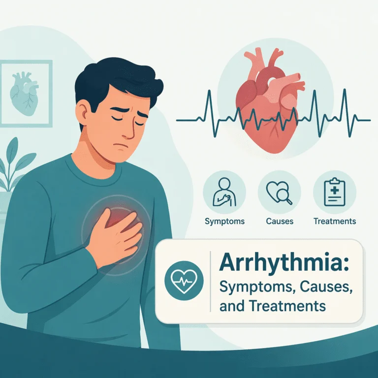
A heartbeat that stumbles, races, or pauses is unsettling, because it happens somewhere you cannot see or control. Most people notice it first as a flutter while lying in bed, a thud that seems to skip a step, or a sudden gallop arriving without warning. Arrhythmia is the term for any rhythm departing from the heart’s normal electrical pattern, and it spans everything from a harmless extra beat to a chaotic rhythm needing emergency care.
Telling the benign from the dangerous is the challenge, because the two can feel identical. This guide covers the heart’s wiring, which symptoms need same-day attention, the workup, and how treatment has changed.
What is an arrhythmia?
Your heart is a muscle governed by an electrical system, and the rhythm at your wrist is a signal traveling a set route at a set speed. It starts in the sinoatrial node, a cluster of cells in the upper right atrium acting as the natural pacemaker, spreads across both atria, pauses at the atrioventricular node so the ventricles can fill, then races down conducting fibers to trigger the contraction that sends blood to the body.
An arrhythmia is any disruption of that sequence. The signal may fire too fast, producing a tachycardia above 100 beats per minute, or too slowly, producing a bradycardia below 60. It may start in the wrong place, as when an irritable patch fires early, or get caught in a loop circling a scar. Not every deviation is a disease; athletes routinely rest in the 40s. What matters is whether the rhythm compromises blood flow or raises stroke risk.
Types of arrhythmia
Cardiologists group arrhythmias by origin and speed. Supraventricular rhythms start above the ventricles and are usually annoying rather than lethal; ventricular rhythms start in the pumping chambers and carry far more weight, because a ventricle in chaos moves no blood. Atrial fibrillation is the most common sustained arrhythmia, affecting several million American adults, and because the fibrillating atria never fully empty, blood pools in the left atrial appendage, clots, and can travel up to trigger the sudden brain-artery blockage responsible for most cardioembolic strokes.
| Type | What happens electrically | Typical concern |
|---|---|---|
| Atrial fibrillation | Chaotic atrial impulses; irregular ventricular response | Clot formation and stroke; fatigue; heart weakening over time |
| Atrial flutter | Organized circular loop in the right atrium, often near 300 per minute | Same stroke risk as fibrillation; highly curable by ablation |
| Supraventricular tachycardia (SVT) | Re-entry circuit at or near the atrioventricular node | Abrupt 150 to 250 bpm episodes; distressing but rarely dangerous |
| Premature ventricular contractions (PVCs) | Early beat from ventricular tissue, followed by a compensatory pause | Usually benign; a very high daily burden can weaken the pump |
| Ventricular tachycardia | Rapid run of beats arising below the atrioventricular node | Can collapse blood pressure and degenerate into fibrillation |
| Ventricular fibrillation | Completely disorganized ventricular activity; no effective output | Cardiac arrest; requires defibrillation within minutes |
| Bradycardia and heart block | Sinus node slowing or interrupted conduction to the ventricles | Fainting and exercise intolerance; often needs a pacemaker |
Symptoms and warning signs
Arrhythmia symptoms are notoriously inconsistent. Roughly a third of people with atrial fibrillation feel nothing and learn of it from a routine exam or a smartwatch alert, while others are floored by the same rhythm. The commonest complaint is palpitations: fluttering, pounding, or a fish flopping in the chest. Extra beats produce the opposite, a momentary void followed by a forceful thud. Other symptoms reflect reduced output: lightheadedness, breathlessness on stairs you normally climb easily, unshakable fatigue, and poor exercise tolerance. Slow rhythms cause foggy thinking and near-fainting instead.
Red flags that mean call 911
Call emergency services immediately for fainting during exertion, chest pain lasting more than a few minutes, severe breathlessness at rest, confusion or slurred speech, one-sided weakness or facial droop, a racing heartbeat that will not settle after 20 to 30 minutes of rest, or any collapse. A fast heart with chest pain and sweating warrants an ambulance, because the cause may be a heart attack triggering the rhythm rather than the reverse.
Causes and risk factors
Arrhythmias arise from a structural heart problem, a chemical or hormonal disturbance, or an inherited ion channel defect. Age is the strongest risk factor for atrial fibrillation, since fibrous scarring in aging atria gives chaotic circuits ideal terrain, and hypertension accelerates that scarring, which is why cardiologists target the sustained pressure overload that gradually reshapes the heart’s chambers. Prior heart attacks, valve disease, cardiomyopathy, and untreated sleep apnea add risk.
Electrolyte disturbances deserve attention because they are common, easily missed, and almost always correctable. Heart cells depend on precise gradients of potassium, magnesium, sodium, and calcium to reset each beat, so vomiting, diarrhea, diuretic use, and alcohol binges can all provoke the potassium imbalance that destabilizes the heart’s electrical recovery phase. Low magnesium is the quiet partner here, because the body cannot retain potassium when magnesium is depleted, and low calcium lengthens repolarization in ways that invite ventricular rhythms.
Thyroid hormone is the other classic reversible trigger, since excess hormone speeds the sinus node and shortens atrial refractory periods. A meaningful share of cases under 60 turn out to reveal an unsuspected overactive thyroid presenting first as a racing heartbeat, and treating the gland often restores normal rhythm without any cardiac drug. Stimulants matter too: high-dose caffeine, nicotine, decongestants, cocaine, and binge drinking, so reliably linked to weekend episodes that clinicians call it holiday heart syndrome.
How arrhythmias are diagnosed
The central difficulty is capture: a rhythm lasting four minutes twice a month will almost never show up on a 10-second office recording, so diagnosis means matching the monitoring window to the symptom frequency.
The 12-lead electrocardiogram, or ECG, is the starting point and is definitive when the arrhythmia is present; it also reveals old scars, chamber enlargement, and conduction blocks even in normal rhythm. For near-daily symptoms, a Holter monitor records every beat for 24 to 48 hours; for weekly symptoms, an event monitor or patch worn two to four weeks works better. Consumer wearables have changed this landscape, catching many silent cases, though any alert needs confirmation on a medical-grade tracing; an implantable loop recorder covers rare events for up to three years. An echocardiogram assesses pumping strength, valve function, and chamber size, showing whether the arrhythmia is standalone or structural. When a cure is the goal, an electrophysiology study threads catheters into the heart to map the signal’s origin.
The blood tests that matter
Laboratory work identifies the reversible causes no ablation can fix. A basic metabolic panel is mandatory, and clinicians add a magnesium level that quietly governs how well potassium stays inside heart cells, since correcting potassium alone often fails. Every new atrial fibrillation workup includes a TSH measurement that reveals an overactive or underactive thyroid. If chest pain accompanied the episode, the team measures a troponin level that signals injured heart muscle. Breathlessness alongside a fast rhythm prompts clinicians to check an NT-proBNP result that reflects rising pressure inside stretched heart chambers. A complete blood count screens for anemia, and kidney function guides anticoagulant dosing.
Treatment options
Treatment answers three questions: does this rhythm threaten the brain, should the rate be slowed or the rhythm restored, and can it be cured. Stroke prevention comes first in atrial fibrillation and flutter. Risk is estimated with the CHA2DS2-VASc score, and most people above the threshold receive a direct oral anticoagulant such as apixaban or rivaroxaban, which have largely replaced warfarin because they need no monitoring and cause fewer brain bleeds. Rate control uses beta-blockers, calcium channel blockers, or occasionally digoxin. Rhythm control uses antiarrhythmic drugs such as flecainide, sotalol, or amiodarone, or electrical cardioversion, a brief synchronized shock under sedation. Catheter ablation destroys the tissue sustaining the arrhythmia and is now first-line for symptomatic SVT, atrial flutter, and many atrial fibrillation cases. Pacemakers correct slow rhythms, while implantable defibrillators terminate life-threatening ventricular rhythms automatically. When a fast rhythm has already weakened the pump and produced the tachycardia-induced heart failure that frequently reverses once rhythm control is achieved, restoring sinus rhythm treats both the symptom and the structure.
Prevention and daily management
Risk-factor modification is not a soft alternative to treatment; trials show it measurably reduces recurrence after ablation. Weight loss of 10 percent or more has one of the strongest effect sizes in the field, and treating sleep apnea with CPAP matters nearly as much, because overnight oxygen drops stretch the atria nightly. Keeping blood pressure below 130/80 protects atrial tissue, and cutting alcohol produces a dose-dependent drop in episodes. Keep caffeine moderate rather than eliminating it, and never stop an anticoagulant yourself.
Living with an arrhythmia: outlook
For most people the outlook is good. PVCs in a structurally normal heart carry a benign prognosis, and SVT is curable in a single procedure. Atrial fibrillation is chronic, but anticoagulation cuts stroke risk substantially, and life expectancy in well-managed patients approaches that of the general population. The psychological burden is underestimated: hypervigilance creates a loop in which anxiety raises adrenaline and adrenaline provokes more extra beats. Naming that loop helps break it, and cardiac rehabilitation addresses it.
Latest scientific advances
A Swiss randomized trial asked whether pulsed field ablation, a newer technique using rapid electrical pulses rather than heat or cold, works as well as cryoballoon ablation, which freezes tissue. All 210 participants with paroxysmal atrial fibrillation received an implantable monitor, so recurrences were caught continuously. Between day 91 and day 365, atrial tachyarrhythmia returned in 37.1 percent of the pulsed field group versus 50.7 percent of the cryoablation group, a gap of 13.6 percentage points meeting criteria for both noninferiority and superiority (Reichlin et al., 2025). What this means for you: if ablation is on the table, the technique used is a fair question to ask.
Must anticoagulation continue forever after a successful ablation? The ALONE-AF trial randomized 840 people at 18 South Korean hospitals who had a stroke risk factor and no recurrence for a full year after ablation, assigning them to stop or continue. Over two years, the combined outcome of stroke, systemic embolism, and major bleeding occurred in 1 patient (0.3 percent) who stopped versus 8 patients (2.2 percent) who continued, and major bleeding hit zero versus 5 patients (1.4 percent) (Kim et al., 2025). What this means for you: stopping may be reasonable for selected people, but that decision belongs with an electrophysiologist reviewing your monitoring data.
The international OPTION trial tested a mechanical alternative in 1,600 ablation patients at elevated stroke risk, comparing left atrial appendage closure, a device sealing the pouch where clots form, against continued anticoagulation. At 36 months, non-procedure-related major or clinically relevant bleeding had occurred in 8.5 percent of the device group versus 18.1 percent of the anticoagulation group, while death, stroke, or systemic embolism was comparable at 5.3 percent and 5.8 percent (Wazni et al., 2024). What this means for you: if bleeding on blood thinners is your obstacle, appendage closure now rests on strong randomized evidence.
Myths and facts
Myth: a skipped beat means something is seriously wrong. Fact: isolated extra beats are extremely common and usually harmless in a structurally normal heart; frequency and accompanying symptoms determine significance.
Myth: if you feel fine, your atrial fibrillation is not dangerous. Fact: silent atrial fibrillation carries the same stroke risk as the symptomatic kind, because prevention depends on your risk profile, not symptoms.
Myth: ablation is a last resort after drugs fail. Fact: guidelines now support ablation as first-line rhythm control for many people with symptomatic paroxysmal atrial fibrillation and for most SVT.
Glossary
| Term | What it means |
|---|---|
| Sinoatrial node | The heart’s natural pacemaker, a cluster of cells in the right atrium that sets the rhythm. |
| Paroxysmal | Occurring in discrete episodes that start and stop on their own, usually within seven days. |
| Catheter ablation | A procedure that destroys the small area of tissue generating or sustaining the arrhythmia. |
| CHA2DS2-VASc | A scoring system that estimates stroke risk in atrial fibrillation and guides anticoagulation. |
| Left atrial appendage | A small pouch off the left atrium where most atrial fibrillation clots form. |
| Repolarization | The electrical reset between beats; disturbances here cause many dangerous rhythms. |
| Electrophysiology study | A catheter-based mapping test that pinpoints exactly where an abnormal rhythm begins. |
Frequently asked questions
What is a normal heart rate, and what is a good resting heart rate with an arrhythmia?
A resting rate of 60 to 100 beats per minute is normal for adults, though values outside that band are not automatically abnormal: endurance athletes often rest in the 40s, while anxiety, fever, or anemia can push a healthy heart above 100. For people with atrial fibrillation on rate-control therapy, cardiologists usually aim for a resting rate under 100 to 110, and a comfortable 60 to 80 suits most patients. Measure it at the same time each day and bring the log to appointments.
What are the most common afib symptoms?
The classic presentation is an irregular, often rapid pulse with no discernible pattern, plus fatigue disproportionate to your activity, breathlessness on modest exertion, and a fluttering chest sensation. Many also report reduced exercise capacity and lightheadedness. Importantly, roughly one in three people has no symptoms at all and is diagnosed through a routine pulse check or smartwatch alert, and that silence does not reduce stroke risk.
Are heart palpitations always a sign of arrhythmia?
No. Palpitations are a symptom rather than a diagnosis, and many have nothing to do with abnormal rhythm. Anxiety, caffeine, nicotine, dehydration, fever, anemia, thyroid overactivity, and decongestants all heighten awareness of the heartbeat. Still, palpitations are the leading symptom of genuine arrhythmias, so recurring episodes deserve at least one ECG and a blood panel. Palpitations with fainting need prompt cardiology assessment.
What does arrhythmia treatment usually involve?
It depends entirely on the rhythm. Some arrhythmias need nothing beyond removing a trigger such as excess alcohol, a stimulant medication, or a low potassium level. Atrial fibrillation treatment typically combines an anticoagulant, a rate-control or rhythm-control strategy, and management of blood pressure, weight, and sleep apnea. SVT and flutter are often cured with a single ablation, while heart block needs a pacemaker.
Can an ECG miss an arrhythmia?
Yes, routinely. A standard ECG records roughly 10 seconds of activity, so an intermittent rhythm must be present at that moment to be caught. A normal tracing alongside convincing episodic symptoms should prompt longer monitoring rather than reassurance: a 24 to 48 hour Holter, a multi-week patch, or a loop recorder for rare events. The ECG still helps, because it reveals underlying scarring.
Sources
- National Heart, Lung, and Blood Institute — Arrhythmias — National Institutes of Health, 2025 — nhlbi.nih.gov
- Centers for Disease Control and Prevention — Atrial Fibrillation — CDC Heart Disease, 2024 — cdc.gov
- American Heart Association — About Arrhythmia — American Heart Association, 2024 — heart.org
- Mayo Clinic — Heart Arrhythmia: Symptoms and Causes — Mayo Clinic, 2024 — mayoclinic.org
- Reichlin T, Kueffer T, Badertscher P, et al. — Pulsed Field or Cryoballoon Ablation for Paroxysmal Atrial Fibrillation — New England Journal of Medicine, 2025 — doi.org
- Kim D, Shim J, Choi JI, et al. — Long-Term Anticoagulation Discontinuation After Catheter Ablation for Atrial Fibrillation: The ALONE-AF Randomized Clinical Trial — JAMA, 2025 — doi.org
- Wazni OM, Saliba WI, Nair DG, et al. — Left Atrial Appendage Closure after Ablation for Atrial Fibrillation — New England Journal of Medicine, 2024 — doi.org
Further reading
- Because narrowed arteries are among the conditions most likely to generate ventricular rhythm problems, read our guide to coronary artery disease and how blocked arteries develop.
- If your palpitations arrive alongside chest tightness during exertion, our explainer describing angina and the chest pain that signals oxygen-starved heart muscle will help you separate the two.
- People whose arrhythmia has begun to affect pumping function should review our article on congestive heart failure and the fluid buildup it produces.
- Calcium plays a direct role in the electrical reset between beats, which makes it worth understanding total calcium results and the repolarization changes they can cause.
Understand your lab results with BloodSense
A surprising share of arrhythmia episodes trace back to something a blood test can reveal. Potassium and magnesium sit at the center of every heartbeat’s electrical reset, and even mildly low values can generate extra beats. TSH identifies the thyroid overactivity that often shows up as unexplained atrial fibrillation, troponin separates a rhythm problem from injured muscle, and BNP or NT-proBNP show the pressure the chambers are under.
Tracking these markers over time beats any single snapshot, because a downward trend in potassium or a slow rise in NT-proBNP often appears before symptoms do. BloodSense turns your lab report into plain-language explanations and the questions worth raising with your cardiologist.