





Atopic Dermatitis: Symptoms, Causes, and Treatments
Few skin conditions are as misunderstood as atopic dermatitis. It is the most common form of eczema, it affects roughly one in ten people in the United States at some point, and it usually announces itself long before a person can describe what they feel — often on the cheeks of a baby a few months old.
What separates it from a passing rash is that it is not really a surface problem. It is a whole-body condition with a skin-shaped symptom, driven by a leaky skin barrier and an immune system tuned to overreact — which is also why it travels in company, through the atopic march.
What is atopic dermatitis?
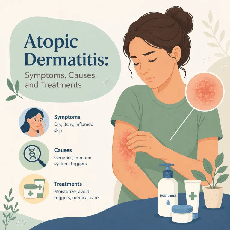
Atopic dermatitis is a chronic, relapsing inflammatory skin disease marked by intense itching, dry skin and recurring patches of inflamed, scaly or weeping rash. It runs in flares rather than a steady state, and between them the skin often still feels rough and tight.
The atopic dermatitis versus eczema question deserves settling early. Eczema is an umbrella term for a family of itchy, inflamed skin conditions — contact dermatitis, dyshidrotic eczema, nummular eczema and seborrheic dermatitis all sit under it. Atopic dermatitis is one specific member, and by far the largest: when someone says “eczema” without qualifying it, they usually mean atopic dermatitis. Readers wanting the wider picture can explore the full eczema guide and its many subtypes.
“Atopic” is the key word. Atopy is an inherited tendency to mount allergic responses to ordinary things — pollen, dust mites, pet dander, certain foods. Atopic dermatitis is that tendency expressed in skin. Most cases begin before age five, though some adults develop it much later.
Symptoms and warning signs
Itch defines the condition. It worsens at night and drives a scratch-itch cycle in which scratching damages the barrier, releasing more inflammatory signals and producing more itch. Sleep loss is often the heaviest burden.
Other atopic dermatitis symptoms include persistent dryness, rash that oozes or crusts during a flare, thickened leathery skin from months of scratching (lichenification), cracking, and darker or lighter patches left after inflammation settles. Some signs need prompt care: yellow crusting, pus or fever suggests bacterial infection, and clusters of small painful blisters on eczematous skin are a medical emergency.
How the rash looks at different ages and on different skin tones
Location shifts with age. In infants the rash favors cheeks, forehead and scalp, sparing the diaper area. In older children it moves into the flexural creases: inner elbows, behind the knees, wrists and neck. In adults it concentrates on hands, eyelids, neck and flexures.
Skin tone changes the picture, and this drives real diagnostic delay. On lighter skin, active disease reads as red or pink. On brown and Black skin, the same inflammation more often appears violet, gray or dark brown, with redness barely visible, and follicular patterns are more common. It also leaves pronounced hyperpigmentation or hypopigmentation lasting months after the flare resolves. Because most medical imagery shows lighter skin, browsing atopic dermatitis pictures can mislead; distribution, thickening and scaling are more reliable than redness.
Causes and risk factors
No single cause explains atopic dermatitis. It emerges where a defective skin barrier, a dysregulated immune response and environmental exposure meet — with genetics loading the dice on the first two.
The barrier side centers on filaggrin, a protein that seals the outer skin layer and breaks down into the compounds that keep skin hydrated. Loss-of-function mutations in the filaggrin gene rank among the strongest genetic risk factors. When filaggrin is deficient, water escapes and allergens, irritants and microbes get in — letting sensitization happen through the skin itself.
The immune side is dominated by type 2 inflammation. Cytokines — chemical messengers immune cells use to coordinate — are overproduced, particularly interleukin-4 and interleukin-13, which inflame skin and weaken the barrier further. Interleukin-31 acts as a direct itch signal along nerve pathways antihistamines never reach, which is why modern drugs target these molecules specifically. Microbiome shifts compound it, with Staphylococcus aureus overgrowing during flares.
Then there is the atopic march. It usually starts with infant eczema, often adds food allergy, and a substantial share of these children later develop asthma and its characteristic airway inflammation. Later still, many go on to develop hay fever and its seasonal allergy symptoms. The march is a tendency, not a destiny, though earlier onset and greater severity raise the odds.
How atopic dermatitis is diagnosed
No single test confirms atopic dermatitis. Diagnosis is clinical: a chronic or relapsing itchy rash, in age-typical locations, with a personal or family history of atopy.
Much of the work is exclusion. Dermatologists routinely rule out psoriasis and its thick, silvery plaques, along with scabies and fungal infection. Patch testing identifies allergic contact dermatitis, which can mimic atopic dermatitis or sit on top of it. It is worth doing when a long-standing case suddenly worsens or stops responding, because an allergy to a preservative, fragrance or topical medication may be the hidden driver.
The blood tests that matter
Bloodwork does not diagnose atopic dermatitis: normal results do not rule it out, and abnormal results do not confirm it. What labs do well is characterize the allergic background, flag complications and set a baseline before systemic treatment.
Allergy workups often include an eosinophil count that reflects allergic activity, which tracks loosely with severity. Total IgE is often elevated, but the healthy range is wide and a high number alone means little. Specific IgE testing answers a narrower question — whether antibodies exist to one pollen, mite, animal or food — yet a positive result signals sensitization, not necessarily a real-world allergy. This is why broad food panels in children often cause harm by triggering needless elimination diets.
A clinician may also order a complete blood count that surveys every major cell line, which captures eosinophils and can reveal infection during a severe flare. Broader panels can measure C-reactive protein as a general marker of systemic inflammation, usually normal here and most informative when unexpectedly high. In atypical or early-onset disease, workups sometimes measure immunoglobulin G and its record of long-term antibody memory to exclude an underlying immunodeficiency. Before oral JAK inhibitors, liver enzymes, kidney function, blood counts and lipids are checked and monitored — safety surveillance, not diagnosis.
Treatment options
Atopic dermatitis treatment works in tiers, escalating only when the foundation proves insufficient. That foundation never disappears: moisturizer stays in the plan even on advanced biologics. Topical corticosteroids remain the workhorse for flares, matched in potency to body site and age, while steroid-sparing topicals cover delicate areas and maintenance. For disease resisting topicals, targeted biologics and oral JAK inhibitors have largely displaced broad immunosuppressants.
| Treatment tier | Examples | What it is for |
|---|---|---|
| Barrier care | Emollients, lukewarm bathing, non-soap cleansers | Daily foundation that cuts water loss and flare frequency |
| Topical anti-inflammatories | Corticosteroids, calcineurin and PDE4 inhibitors, topical JAK inhibitors, tapinarof | Treating active flares and, used proactively, preventing the next one |
| Adjuncts | Wet wraps, dilute bleach baths, antibiotics for confirmed infection | Calming severe flares and bacterial overgrowth |
| Phototherapy | Narrowband UVB in a clinic | Widespread disease when topicals fall short |
| Biologics | Dupilumab, tralokinumab, lebrikizumab, nemolizumab | Moderate-to-severe disease; injected, cytokine-targeted, no routine monitoring |
| Oral JAK inhibitors | Upadacitinib, abrocitinib | Moderate-to-severe disease needing fast itch control; tablets, monitored |
| Conventional immunosuppressants | Cyclosporine, methotrexate, azathioprine, mycophenolate | Older off-label options, reserved for access or cost constraints |
Oral corticosteroids warrant caution: they work fast but are advised against routinely, because the disease reliably rebounds, often worse, once they stop.
Triggers, skincare and daily management
Trigger management is personal — identify your own rather than adopting a generic list. Frequent culprits include soaps and fragrance, wool and rough synthetics, sweat, low humidity, dust mites and stress.
Routine beats products. Short lukewarm showers, a gentle non-soap cleanser, patting dry, and moisturizing within minutes of towel-drying while skin is damp accomplish more than most people expect. Thick ointments outperform lotions, which are largely water and sting broken skin. Sedating antihistamines may aid sleep without treating the itch itself, which is not primarily histamine-driven.
On diet, food allergy is genuinely more common here, but removing foods rarely clears skin and unsupervised elimination risks nutritional deficiency. Some patients also monitor vitamin D levels and their connection to immune regulation, though supplementation is not itself a treatment.
Living with atopic dermatitis: outlook
There is no cure, but the outlook has improved markedly. Many children clear substantially by their teens; others follow a course that quiets and periodically returns. Adults with persistent disease now have targeted options that can reach near-clear skin, an unrealistic goal before 2017.
The burden extends past skin. Chronic itch and broken sleep are linked to higher rates of anxiety and depression, reduced school and work performance, and social withdrawal — effects underestimated in short appointments and worth raising, because they influence how aggressively treatment should escalate.
Latest scientific advances
The most informative recent trial compared two modern drugs directly rather than against placebo. The Level Up study randomly assigned adolescents and adults with moderate-to-severe disease to either upadacitinib, an oral JAK inhibitor (a tablet blocking Janus kinase enzymes that relay inflammatory signals inside cells), or dupilumab, an injected biologic blocking interleukin-4 and interleukin-13 signaling. At week 16, 19.9% on upadacitinib hit the demanding combined target of at least 90% improvement in eczema severity plus little-to-no itch, versus 8.9% on dupilumab (P < 0.001), with itch differences appearing by day 2 (Silverberg et al., 2025). What this means for you: if rapid, near-complete itch relief is the priority, an oral JAK inhibitor may outperform a biologic, but it requires ongoing blood monitoring — a genuine trade-off to weigh with a specialist.
A 2025 review in the Annals of Allergy, Asthma & Immunology mapped how far the treatment landscape has widened, drawing on peer-reviewed studies, conference abstracts and regulatory filings. Dupilumab is now approved from age 6 months; tralokinumab, lebrikizumab and nemolizumab — the last targeting the interleukin-31 receptor behind non-histamine itch — from age 12; and the non-steroidal topicals roflumilast and tapinarof from ages 6 and 2 (Gallagher et al., 2025). What this means for you: if you were told years ago that steroids and moisturizer were the only options, that advice is out of date, and age is far less of a barrier to targeted treatment.
Pediatric guidance moved in parallel. A 2025 clinical report in Pediatrics reviewed skin-directed management in children, noting that atopic dermatitis affects 20% to 25% of children and reflects an interplay between a defective barrier, immune dysfunction and the skin microbiome. It reaffirmed topical corticosteroids plus moisturization as standard care, found proactive treatment with corticosteroids or calcineurin inhibitors reduces flares, and stressed keeping regimens simple (Schoch et al., 2025). What this means for you: a simple routine you actually follow beats an elaborate one you abandon, so asking a clinician to strip a complicated plan back to essentials is a legitimate request.
Myths and facts
Myth: atopic dermatitis is contagious. Fact: it cannot pass by touch, shared towels or swimming pools. Genetics and immune function drive it, not an organism.
Myth: it reflects poor hygiene. Fact: the reverse is closer — harsh, frequent soap washing strips the barrier and worsens it.
Myth: topical steroids are dangerous. Fact: at appropriate potency and duration they are effective, and steroid fear typically produces longer flares and more medication use.
Myth: moisturizers are cosmetic. Fact: they are treatment, reducing flare frequency and medication needed.
Glossary
| Term | What it means |
|---|---|
| Atopy | An inherited tendency to mount allergic responses to ordinary substances |
| Atopic march | Progression from infant eczema to food allergy, asthma and hay fever |
| Filaggrin | A skin protein that seals the barrier and retains moisture; faulty versions are a major risk factor |
| Cytokine | A chemical messenger immune cells use; interleukin-4, -13 and -31 are central here |
| Lichenification | Thickened, leathery skin with exaggerated lines, from prolonged scratching |
| Pruritus | The medical term for itch |
| Emollient | A moisturizing ointment or cream that limits water loss |
| Biologic | An injected antibody drug blocking one specific immune signal, not immunity broadly |
| JAK inhibitor | A drug blocking Janus kinase enzymes to interrupt signaling inside cells |
Frequently asked questions
What is the difference between atopic dermatitis and eczema?
Eczema is the umbrella category for itchy, inflamed skin conditions, including contact dermatitis, dyshidrotic eczema and seborrheic dermatitis. Atopic dermatitis is one specific type within that group and by far the most common, which is why the terms get used interchangeably. The distinction is that atopic dermatitis is tied to atopy, the inherited allergic tendency, and usually begins in childhood alongside a family history of allergy or asthma.
Is atopic dermatitis curable?
There is no cure, but that sounds bleaker than it is. Control is realistic, and for many people now means long stretches of clear or near-clear skin. Children frequently improve as they grow, and adults with persistent disease have biologics and JAK inhibitors unavailable a decade ago. Treatment aims to reduce flares and protect sleep and quality of life rather than erase the underlying tendency.
Is atopic dermatitis contagious?
No. It cannot be caught from or given to anyone through skin contact, shared towels, bedding or swimming pools. It arises from inherited differences in the skin barrier plus an immune system prone to allergic responses. One caveat causes confusion: eczematous skin is vulnerable to infection, and those secondary infections can be contagious. That is an infection sitting on top of atopic dermatitis, not the condition spreading.
Is atopic dermatitis an autoimmune disease?
Not in the strict sense. In autoimmune disease the immune system attacks the body’s own tissues. In atopic dermatitis it overreacts to external substances such as allergens and microbes while a weakened barrier lets them in, so it is better described as an immune-mediated inflammatory disease. The distinction matters, because treatment damps specific allergic signaling pathways and repairs the barrier rather than applying classic autoimmune suppression.
What causes atopic dermatitis?
Three factors combine. A genetically weakened skin barrier, often involving filaggrin gene mutations, lets moisture out and irritants, allergens and bacteria in. An immune system tuned toward type 2 inflammation overproduces cytokines including interleukin-4, interleukin-13 and interleukin-31, driving inflammation and itch. Environmental exposure — dry air, hard water, stress and microbiome shifts such as Staphylococcus aureus overgrowth — then sustains flares. No single factor explains the condition alone.
Does atopic dermatitis go away?
Often it improves substantially. A large share of children see symptoms fade by adolescence, particularly those with milder, later-onset disease. Improvement is not disappearance, though: many retain sensitive, dry skin that flares under the right conditions, some carry active disease into adulthood, and a subset develops it first as adults. Earlier onset, greater severity and coexisting asthma all make persistence more likely.
Sources
- NIAMS — Atopic Dermatitis — National Institutes of Health, 2024 — niams.nih.gov
- MedlinePlus — Eczema (Atopic Dermatitis) — U.S. National Library of Medicine, 2024 — medlineplus.gov
- Mayo Clinic — Atopic dermatitis (eczema) — Mayo Clinic, 2024 — mayoclinic.org
- American Academy of Dermatology — Atopic dermatitis overview — AAD, 2024 — aad.org
- National Eczema Association — Atopic Dermatitis — National Eczema Association, 2024 — nationaleczema.org
- Cleveland Clinic — Atopic Dermatitis — Cleveland Clinic, 2023 — clevelandclinic.org
- Silverberg JI, et al. — Upadacitinib versus dupilumab in moderate-to-severe atopic dermatitis: Level Up week 16 results — British Journal of Dermatology, 2025 — doi.org
- Gallagher K, et al. — New treatments in atopic dermatitis update — Annals of Allergy, Asthma & Immunology, 2025 — doi.org
- Schoch JJ, et al. — Atopic Dermatitis: Update on Skin-Directed Management — Pediatrics, 2025 — doi.org
Further reading
- Anyone comparing subtypes should read the complete eczema guide covering every variant.
- Parents tracking the atopic march will want the asthma overview explaining symptoms and treatments.
- Anyone reviewing an allergy panel should understand basophils and the part they play in allergic reactions.
- Readers curious about mucosal immunity can examine immunoglobulin A and its role in barrier defenses.
- Those exploring the inflammation connection may value the analysis linking gut health, skin and inflammation markers.
Understand your lab results with BloodSense
Atopic dermatitis is diagnosed by a clinician looking at your skin, not by a blood test — yet labs surround the condition at every stage. An eosinophil count and total IgE sketch the allergic background, a complete blood count can reveal infection during a flare, and C-reactive protein shows whether inflammation reaches beyond the skin. Once you start an oral JAK inhibitor, liver enzymes, kidney function and blood counts become an ongoing part of care.
The difficulty is that these numbers arrive as a dense PDF of reference ranges with no narrative. BloodSense reads your results and explains them in plain language — which markers sit outside range, what patterns they form, and what to raise at your next appointment.