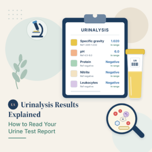
Seeing a note about crystals in urine on a lab report is unsettling, but in most cases it describes a normal chemical event rather than a disease. Urine is a concentrated solution of minerals, salts and waste products, and when that solution becomes crowded enough, some of those substances drop out of liquid form and settle as visible particles. A laboratory technician looking through a microscope names the shape they see, and that name lands on your report.
In this article you will learn which crystal types are common and usually harmless, which ones always deserve a follow-up appointment, why urine acidity and the time your sample spent on a bench change the result, and how crystals relate to kidney stones. You will also find a crystal-by-crystal reference table, a plain-language glossary and answers to the questions patients ask most.
What a crystal in your urine sample actually is
Your kidneys filter roughly 50 gallons of fluid every day and return most of the water to your bloodstream. What leaves your body is a compact solution carrying calcium, oxalate, phosphate, uric acid, ammonium and dozens of other dissolved substances. Chemistry sets a ceiling on how much of each one can stay dissolved. Push past that ceiling and molecules start locking together into an organized solid, which is what a crystal is.
Why supersaturation matters more than quantity
Laboratories describe crystals qualitatively, using words like rare, few, moderate or many, rather than a precise count. That is deliberate. The number seen on one slide reflects the concentration of that single sample, not a stable property of your body. A dehydrated morning sample from a healthy person can contain plenty of crystals, while the same person tested two hours after drinking a liter of water may show none at all.
What the microscope adds to the dipstick
Crystals only appear on the microscopic portion of a urinalysis, never on the chemical dipstick. If you want to see how the two halves of the test fit together, read our guide to urine microscopy results. The microscopic exam also reports cells, bacteria and cylinder-shaped protein structures, and clinicians read all of those findings together rather than one line at a time. Anyone puzzled by the cylinder-shaped entries can consult our explanation of urinary casts and what they indicate.
The crystals that are usually harmless
Three groups account for the overwhelming majority of crystals reported in routine samples, and all three are frequently found in people with no kidney problem whatsoever.
Calcium oxalate
Calcium oxalate is the crystal most often named on American lab reports. It forms across a wide range of urine acidity, which is why it turns up so consistently. Two shapes exist: a square envelope form (the dihydrate) and a spindle or dumbbell form (the monohydrate). The envelope form is the everyday version and is strongly influenced by diet, since spinach, rhubarb, beets, nuts and strong tea all deliver oxalate. Occasional calcium oxalate crystals in urine carry no meaning on their own. Large numbers, especially of the spindle-shaped monohydrate variety, are the finding a clinician looks at more carefully.
Amorphous urates and amorphous phosphates
Amorphous simply means shapeless. These appear as a grainy pink or white haze rather than a defined geometric figure, and they are the single most common cause of a cloudy sample that has been sitting in a refrigerator. Urates form in acidic urine, phosphates in alkaline urine, and neither has any clinical significance in a healthy person. They are essentially an artifact of the sample cooling down.
Triple phosphate (struvite)
Triple phosphate crystals have a distinctive coffin-lid shape and form only in alkaline urine. In a fresh sample from a person with no symptoms they can be entirely benign. Their significance changes when they appear alongside bacteria, white blood cells or a positive nitrite result, because certain bacteria raise urine pH by splitting urea and thereby create the exact conditions struvite needs. Anyone with those combined findings should investigate a possible urinary tract infection and its urine test pattern. The chemical marker that flags the same bacteria is explained in our page on nitrite in a urine test.
The crystals that always need follow-up
A short list of crystal types is considered abnormal in any quantity, in any person, at any age. Finding one of them is not an emergency, but it does mean the result should be discussed with a clinician rather than filed away.
Cystine
Cystine forms flat six-sided plates and signals cystinuria, an inherited condition in which the kidney fails to reabsorb an amino acid and instead dumps it into the urine. Cystine dissolves poorly, so people with the condition form stones repeatedly, often starting in childhood or early adulthood. Because it is genetic, a single positive finding changes lifelong management: high fluid intake, urine alkalinization and periodic imaging.
Tyrosine and leucine
These two appear as fine needles and as oily-looking spheres with concentric rings. Both point toward severe liver dysfunction or a rare inherited metabolic disorder, because a healthy liver processes these amino acids rather than letting them spill into urine. They are genuinely uncommon, and when reported they usually accompany other abnormal findings such as bilirubin. Readers who see that marker on their own report can review our guide to bilirubin in a urine sample.
Drug crystals
Certain medications crystallize in the renal tubules when urine is concentrated or when the dose is high. Sulfonamide antibiotics are the classic example, and acyclovir, indinavir, methotrexate and high-dose intravenous vitamin C can do the same. Drug crystals matter because they can physically obstruct the kidney and cause acute injury. They are reported so that the prescribing clinician can adjust fluids, dose or the drug itself. Anyone reviewing a report that also covers medications may want to compare it with our overview of urine drug screen results.
A crystal-by-crystal reference table
This table pairs each crystal type with the urine acidity it prefers and the way clinicians usually interpret it. Urine pH runs from about 4.5 to 8.0; values below 7 are acidic and values above 7 are alkaline.
| Crystal type | Urine pH where it forms | Usual significance |
|---|---|---|
| Calcium oxalate | Any pH, most often acidic to neutral | Very common and usually harmless; heavy amounts raise stone risk |
| Amorphous urates | Acidic (below 6) | No clinical meaning; often a cooling artifact |
| Amorphous phosphates | Alkaline (above 7) | No clinical meaning; common cause of a cloudy sample |
| Uric acid | Acidic (5.0 to 5.5) | Often benign; linked to gout, dehydration and high cell turnover |
| Triple phosphate (struvite) | Alkaline (above 7) | Benign alone; suspicious for infection with bacteria or white cells |
| Calcium phosphate | Alkaline (above 7) | Usually benign; persistent amounts prompt a metabolic check |
| Ammonium biurate | Alkaline | Almost always from an old or poorly stored sample |
| Cystine | Acidic | Always abnormal; indicates inherited cystinuria |
| Tyrosine | Acidic | Always abnormal; suggests liver or metabolic disease |
| Leucine | Acidic | Always abnormal; suggests liver or metabolic disease |
| Drug crystals (sulfa, acyclovir, indinavir) | Varies with the drug | Always reported; medication or hydration may need adjusting |
Why urine pH and sample handling decide what forms
Two variables explain most of the variation between one urinalysis and the next, and neither of them is a disease.
Acidity sets the menu
Each substance has an acidity range in which it stays dissolved and a range in which it precipitates. Uric acid needs acidic urine, so a person eating a heavy meat diet and drinking little water creates ideal conditions for it. Phosphate and struvite need alkaline urine, which is why they appear after vegetarian meals, in some infections and in people whose kidneys have trouble excreting acid. Understanding the number on your own report starts with our page on urine pH test results.
Time and temperature manufacture crystals
This is the single most under-appreciated fact about crystalluria. A sample that sits at room temperature or in a refrigerator for hours before analysis will grow crystals that were never present in the bladder. Cooling reduces solubility, bacteria may multiply and shift the pH upward, and dissolved carbon dioxide escapes. Laboratories therefore want samples examined within about two hours. If your report shows abundant amorphous material and nothing else abnormal, delayed processing is the most likely explanation. Concentration itself is measured separately, and our guide to urine specific gravity results shows how to tell a concentrated sample from a dilute one.
How crystals relate to kidney stones
Every kidney stone begins as a crystal, but the overwhelming majority of crystals never become a stone. The step that separates the two is retention: a crystal has to stick to the kidney lining and stay long enough for more material to deposit on it. Most crystals are simply washed out.
What actually raises stone risk
Risk climbs when the same crystal type appears repeatedly across several samples, when a person is chronically dehydrated, and when a 24-hour urine collection shows high calcium, high oxalate, high uric acid or low citrate. Citrate is the body’s natural crystallization inhibitor, and low levels remove a protective brake. Anyone with a personal or family history should read our full guide to kidney stone causes and prevention.
The uric acid and gout connection
Uric acid crystals in urine and gout share the same chemistry, though the crystals settle in different places: joints in gout, the urinary tract in stone disease. Persistently acidic urine is the decisive factor, and it is common in people with metabolic syndrome or type 2 diabetes. Two BloodSense pages cover the blood-side markers, our page on uric acid blood test results and our overview of gout symptoms and uric acid testing.
When to see a doctor
Book an appointment promptly if crystals appear together with severe flank or side pain, fever or chills, visible blood in the urine, persistent burning during urination, nausea and vomiting alongside pain, or a known history of stones. Book a routine appointment if a report names cystine, tyrosine, leucine or a drug crystal, if the same crystal type recurs on several tests, or if you have chronic kidney disease. Blood in a sample deserves its own attention, and our page on blood in urine test results explains the workup.
Practical steps that reduce crystal formation
The most effective measure by a wide margin is fluid intake. The National Institute of Diabetes and Digestive and Kidney Diseases recommends drinking enough liquid, mainly water, as the single most important step for stone prevention, and describes the approach in its guidance on eating, diet and nutrition for kidney stones. Diluting urine lowers the concentration of every dissolved substance at once, which is why it works regardless of crystal type.
Beyond fluids, the useful measures depend on which crystal was found. Reducing sodium lowers the amount of calcium the kidney excretes. Keeping dietary calcium at a normal level is protective rather than harmful, because calcium binds oxalate in the gut before it can reach the kidney. Moderating animal protein reduces both uric acid production and urinary acidity. Citrus fruit raises citrate. None of these steps should replace a conversation with your clinician, and none of them should be started on the basis of a single crystal finding.
Latest scientific advances
Research published over the last three years has sharpened what urine crystals can and cannot tell us. According to articles indexed in PubMed, the most useful developments concern prediction, rare disease detection and prevention.
A Japanese team studied 164 people who had already formed at least one kidney stone and deliberately cooled their urine samples to force crystallization. Before cooling, crystals were visible in about one sample in ten; after cooling, in three quarters of them. One particular crystal, brushite, showed up far more often in patients whose stones had come back quickly than in first-time stone formers. What this means for you: an inexpensive tweak to a routine test may one day flag who needs intensive prevention, though this was a single-center study and needs confirmation elsewhere before it changes practice.
A 2026 case report showed how much a microscope can reveal when the count is extreme. A woman with stones dating back to childhood had more than 200 calcium oxalate monohydrate crystals per cubic millimeter, a density that pointed her team toward primary hyperoxaluria type 3, an inherited disorder confirmed afterward by genetic testing. What this means for you: an unusually heavy, persistent crystal load is worth investigating rather than dismissing, even when standard 24-hour urine values look acceptable. A single case report is the weakest form of evidence, but it illustrates a real diagnostic route.
A retrospective study of 41 children with cystic fibrosis found calcium oxalate crystals in roughly a third of them and small kidney stones in about one in five, usually with normal kidney function and no symptoms. What this means for you: in some chronic conditions, periodic urine microscopy can catch a problem early, and crystals in a child with a known genetic condition warrant a closer look. This was a small single-center review, so the exact percentages should not be generalized.
On prevention, an umbrella review that pooled 17 earlier meta-analyses examined 46 possible risk factors for kidney stones. Central obesity, type 2 diabetes, gout, high dietary sodium, fructose intake and hot climates were linked to higher risk, while dietary calcium, coffee, dietary fiber and the DASH eating pattern were linked to lower risk. What this means for you: the long-standing worry that dietary calcium causes stones is not supported. An umbrella review summarizes other reviews rather than patients directly, so it shows consistent patterns rather than proof of cause.
Finally, a 2026 systematic review of 23 studies covering more than 78,000 people found that uricosuric drugs, which treat gout by pushing uric acid out through the kidneys, come with a modestly higher rate of stones than other gout medications, affecting roughly three people in a hundred. What this means for you: if you take a gout medication and your report shows uric acid crystals, mention it at your next visit, since fluid intake or urine alkalinization may be adjusted. The authors rated the evidence quality as low to moderate.
Glossary
| Term | Definition |
|---|---|
| Crystalluria | The medical term for the presence of crystals in a urine sample. It describes a finding, not a diagnosis. |
| Supersaturation | A state in which urine holds more of a substance than it can normally keep dissolved. It is the trigger for crystal formation. |
| Amorphous | Without a defined shape. Amorphous urates and phosphates look like fine grains rather than geometric figures. |
| Struvite | Another name for triple phosphate, a coffin-lid shaped crystal made of magnesium, ammonium and phosphate that forms in alkaline urine. |
| Cystinuria | An inherited condition in which the kidney leaks the amino acid cystine into urine, causing repeated stones from a young age. |
| Oxalate | A compound made by the liver and also absorbed from foods such as spinach, nuts and beets. It binds calcium to form the most common crystal type. |
| Citrate | A natural inhibitor of crystallization found in urine. Low urinary citrate removes a protective brake against stone formation. |
| Urine pH | A measure of how acidic or alkaline urine is, on a scale from roughly 4.5 to 8.0. It determines which crystals can form. |
| Nephrolithiasis | The medical term for kidney stones, meaning stone formation inside the kidney. |
| 24-hour urine collection | A test in which all urine passed over a full day is collected, allowing measurement of daily calcium, oxalate, uric acid and citrate output. |
FAQ
What causes crystals in urine?
The direct cause is concentration. When urine carries more of a mineral or waste product than it can keep dissolved, that substance settles out as a solid. The most frequent everyday triggers are drinking too little fluid, a diet heavy in oxalate-rich foods or animal protein, and urine acidity that favors a particular substance. Less common causes include urinary infections that make urine alkaline, certain medications, inherited metabolic conditions and liver disease. Sample handling matters too: a specimen that sits for several hours before analysis often grows crystals that were not present when you produced it.
Are calcium oxalate crystals in urine dangerous?
In most cases, no. Calcium oxalate is the crystal most often reported in healthy adults and a small number carries no clinical weight on its own. The picture changes when large numbers appear repeatedly across several samples, when the spindle-shaped monohydrate form dominates, or when the finding accompanies flank pain, blood in the urine or a history of stones. In those situations a clinician may order a 24-hour urine collection to measure how much calcium, oxalate and citrate you actually excrete in a day.
Is there a normal range for crystals in urine?
There is no numeric reference range in the way there is for glucose or creatinine. Laboratories grade crystals descriptively as rare, few, moderate or many, and interpretation depends on which type was seen. Common types such as calcium oxalate, amorphous urates and phosphates are accepted as normal findings in small to moderate amounts. Types such as cystine, tyrosine, leucine and drug crystals are considered abnormal in any quantity and prompt further evaluation regardless of how many were counted.
Which foods should I avoid if I have crystals in my urine?
Broad dietary restriction is rarely the right answer, and the useful changes depend on which crystal was found. For calcium oxalate, moderating very high oxalate foods such as spinach, rhubarb, beets, almonds and strong black tea can help, while keeping normal dietary calcium is protective rather than harmful. For uric acid, limiting red meat, organ meats and shellfish is more relevant. Cutting sodium and sugary drinks helps across most crystal types. Discuss any restriction with your clinician or a dietitian before starting it.
Do crystals in urine during pregnancy mean something is wrong?
Usually not. Pregnancy changes fluid balance, urine concentration and urine acidity, so crystals are a common incidental finding on the routine urinalysis done at prenatal visits. What matters is the rest of the report. Crystals alongside bacteria, white blood cells or a positive nitrite result may point to a urinary infection, which is treated promptly in pregnancy. Crystals alongside protein prompt a separate evaluation. Report any burning, urgency, fever or back pain to your obstetric team rather than waiting for the next scheduled visit.
Can medications cause crystals in urine?
Yes. Sulfonamide antibiotics are the best-known example, and acyclovir, indinavir, methotrexate, triamterene and high-dose intravenous vitamin C can all crystallize in the kidney when urine is concentrated. This is why drug crystals are always reported: the concern is that they can physically block the small tubes inside the kidney. The usual responses are increasing fluid intake, adjusting the dose, or shifting urine acidity. Never stop a prescribed medication on your own because of a crystal finding; bring the report to the prescribing clinician instead.
Sources
- Cleveland Clinic — Crystals in Urine: Types, Causes, Symptoms & Treatment — Cleveland Clinic Health Library, reviewed 2021 — my.clevelandclinic.org
- MedlinePlus, National Library of Medicine — Urinalysis — MedlinePlus Health Topic, updated 2024 — medlineplus.gov
- National Institute of Diabetes and Digestive and Kidney Diseases — Eating, Diet, & Nutrition for Kidney Stones — NIDDK, reviewed 2017 — niddk.nih.gov
- Mayo Clinic — Kidney stones: Symptoms and causes — Mayo Clinic, 2024 — mayoclinic.org
- Tanaka Y, Tsujino I, Yoshikawa HY, et al. — Cooling-induced brushite crystallization in urine as a predictive risk marker for calcium kidney stone recurrence — Urolithiasis, 2025 — doi.org/10.1007/s00240-025-01820-2
- Santos D, Lança MB, Navarro D, et al. — Urine microscopy revealing a metabolic disorder: a case report — BMC Nephrology, 2026 — doi.org/10.1186/s12882-026-04946-6
- Asfuroglu P, Asfuroglu A — Urinary System Involvement in Children With Cystic Fibrosis: A Single-Center Retrospective Cohort — Pediatric Pulmonology, 2026 — doi.org/10.1002/ppul.71551
- Ma Y, Cheng C, Jian Z, et al. — Risk factors for nephrolithiasis formation: an umbrella review — International Journal of Surgery, 2024 — doi.org/10.1097/JS9.0000000000001719
- Dahanayake C, Bajpai R, Lambie M, et al. — Incidence and risk of nephrolithiasis with uricosuric therapies for the treatment of gout: a systematic review and meta-analysis — Rheumatology (Oxford), 2026 — doi.org/10.1093/rheumatology/keaf577
Further reading
- Check the acidity value that decides which crystals can form by reading our urine pH test guide.
- Explore the rest of the microscopic exam with our urine microscopy results guide.
- Compare your crystal finding with the marker behind gout and stone risk in our uric acid in urine guide.
- Understand how a suspected infection is confirmed by reading our urine culture results guide.
- Review the full picture of stone formation and prevention in our kidney stones guide.
Understand your lab results with BloodSense
A line about crystals rarely means much on its own, but it means a great deal alongside urine pH, specific gravity, blood, bacteria and your kidney function results. BloodSense reads your whole urinalysis together and explains in plain language which findings are routine and which deserve a conversation with your clinician. It helps you understand your report; it does not diagnose you and it does not replace your doctor.