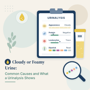
Urinalysis results describe what a lab found when it looked at your urine, tested it with a chemical dipstick and examined it under a microscope. A typical report lists more than 15 items, from color and specific gravity to white blood cells and crystals, and most of them should read negative, normal or within a narrow range. A single abnormal line rarely means much on its own; what matters is the pattern, your symptoms and whether the sample was collected cleanly.
In this article you’ll learn what each part of a urinalysis checks, the normal values for every common item, how doctors read typical patterns such as infection or blood in the urine, how to give a reliable sample and when a result needs follow-up.
What a urinalysis checks
A urinalysis is one of the most common lab tests. Mayo Clinic explains that it is used for routine checkups, pregnancy visits and before surgery, to look into symptoms such as painful or frequent urination, back pain or blood in the urine, and to monitor known conditions such as kidney disease or urinary tract infections. According to the MedlinePlus Medical Encyclopedia, the lab examines the sample in three ways.
- Visual exam: the color and clarity of the urine.
- Dipstick (chemical) test: a strip with small pads that change color to show acidity (pH), concentration, protein, sugar, ketones, bilirubin, blood and signs of infection.
- Microscopic exam: a look at cells, crystals, casts, mucus and germs such as bacteria or yeast.
Not every report includes all three parts. Some labs perform the microscopic exam only when the dipstick is abnormal or when it is specifically requested. If your urinalysis results include a separate section of counts per field, you can see what those numbers mean by reading our urine microscopy guide.
Normal urinalysis results at a glance
The values below come from Cleveland Clinic. Reference ranges vary slightly between laboratories, so always compare your report with the range your lab prints next to each result.
| Item | Normalbefund | What an abnormal result can point to |
|---|---|---|
| Farbe | Pale to dark yellow | Foods, medicines, concentration or blood |
| Clarity | Clear or slightly cloudy | Infection, cells, crystals or mucus |
| Spezifisches Gewicht | 1.005 to 1.030 | Dehydration (high) or very dilute urine (low) |
| pH | 5.0 to 8.0 | Infection, kidney problems, ketoacidosis or diarrhea |
| Protein | Negative or trace | Kidney damage, heart failure, heavy exercise or dehydration |
| Glukose | Negative or trace | Diabetes or gestational diabetes |
| Ketone | Negative or trace | Fasting, low-carbohydrate diets or diabetic ketoacidosis |
| Bilirubin | Negativ | Liver or bile duct problems |
| Blood (hemoglobin) | Negative or trace | Infection, stones, injury or, rarely, cancer |
| Nitrit | Negativ | Bacteria that cause urinary tract infections |
| Leukozytenesterase | Negative or trace | White blood cells from inflammation or infection |
| Rote Blutkörperchen | 0 to 3 per high-power field | Bleeding anywhere in the urinary tract |
| Weiße Blutkörperchen | 0 to 5 per high-power field | Infection or inflammation |
| Casts | 0 per low-power field | Some types point to kidney disease |
| Bacteria and yeast | Keiner | Infection or a contaminated sample |
MedlinePlus adds that glucose, ketones, protein and bilirubin are usually not detectable in normal urine, and that hemoglobin, nitrites, red blood cells and white blood cells are not normally found. Its encyclopedia page on red cells in urine gives the normal result as 4 red blood cells per high-power field or less, a reminder that cutoffs differ slightly between sources.
Reading the visual exam: color and clarity
Normal urine ranges from almost colorless to dark yellow. Darker urine is usually more concentrated, for example first thing in the morning or when you have not had much to drink. MedlinePlus notes that beets and blackberries can turn urine red, and Mayo Clinic adds rhubarb. Several medicines can also change the color without signaling disease, including iron supplements, levodopa, nitrofurantoin, phenazopyridine, phenytoin, riboflavin and triamterene. To decode an unusual shade, read our urine color guide.
Mayo Clinic explains that cloudiness or an unusual odor can suggest a problem such as infection, that protein can make urine look foamy and that blood can make it look red or brown. For the most common reasons urine looks hazy or bubbly, explore our cloudy and foamy urine guide.
Reading the dipstick results
The dipstick section usually makes up most of your urinalysis results. Each pad is read after a set time and reported as negative, trace, a number of plus signs or a concentration.
Specific gravity and pH
Specific gravity shows how concentrated your urine is. The MedlinePlus Medical Encyclopedia gives a normal range of 1.005 to 1.030. High values are most often linked to dehydration, and less often to heart failure or glucose in the urine; low values can come from drinking a lot of fluid, diabetes insipidus or kidney problems. A very dilute sample can also hide abnormal findings. Learn more by reading our Leitfaden zum spezifischen Gewicht des Urins. Urine pH reflects acidity; Cleveland Clinic notes that a high pH can point to kidney problems or a urinary tract infection, while a low pH can occur with diabetic ketoacidosis or diarrhea.
Protein
Healthy kidneys keep almost all protein in the blood. Cleveland Clinic links protein in the urine to kidney damage and heart failure, but also to temporary causes such as heavy exercise and dehydration. Your doctor may repeat the test or order a urine albumin-to-creatinine ratio to measure it more precisely. Review the main causes by reading our urine protein guide.
Glucose and ketones
Sugar spills into urine when blood sugar rises above what the kidneys can hold back, so glucose in the urine is often linked to diabetes or gestational diabetes. Ketones appear when the body burns fat for fuel, which can happen with fasting, low-carbohydrate diets or, more seriously, diabetic ketoacidosis. A positive urine glucose may lead to a blood glucose or A1C test; learn more by reading our urine glucose guide.
Blut
The blood pad reacts to hemoglobin from red blood cells. Cleveland Clinic lists infection, damage to the urinary tract, high blood pressure and cancer among possible causes. A positive dipstick is usually checked by counting red blood cells under the microscope, and menstrual blood can also turn the pad positive, as Cleveland Clinic notes. To see how doctors assess it, read our blood in urine guide.
Leukocyte esterase and nitrite
These two pads are the dipstick’s infection markers. Leukocyte esterase is an enzyme released by white blood cells, and nitrite is made when certain bacteria convert nitrate in urine. Cleveland Clinic notes that either result, or a raised white cell count, often points to inflammation or infection, most commonly a urinary tract infection. Nitrite can stay negative in some infections, because not all bacteria make it. Learn how the first is read by opening our leukocyte esterase guide. The second is covered when you read our urine nitrite guide.
Bilirubin and urobilinogen
Bilirubin is a yellow pigment made when red blood cells break down. It is normally negative in urine, and a positive result may suggest a liver or bile duct problem, according to Cleveland Clinic. Urobilinogen, a related breakdown product, is normally present in small amounts.
Reading the microscopic exam
In the microscopic part of your urinalysis results, the lab counts what it sees per high-power field (HPF) or low-power field (LPF). Mayo Clinic and Cleveland Clinic describe the main findings.
- Red blood cells: more than about 3 to 4 per HPF means blood that may not be visible to the eye, which can come from the kidneys, bladder or anywhere along the urinary tract. MedlinePlus lists infections, stones, kidney inflammation, prostate problems and, less often, cancer as causes.
- White blood cells: more than about 5 per HPF usually signals infection or inflammation.
- Bacteria, yeast or parasites: may indicate infection, but can also come from skin contamination.
- Epithelial cells: squamous cells usually mean the sample was contaminated by skin cells; high numbers of other types can reflect infection, inflammation or, rarely, cancer.
- Casts: tube-shaped particles formed in the kidneys; some types are harmless, while others point to kidney problems.
- Crystals: certain types may suggest kidney stones.
Common patterns in urinalysis results
Doctors rarely diagnose anything from a urinalysis alone, as both Cleveland Clinic and Mayo Clinic stress. They read the results as a pattern and decide what to check next.
| Befundmuster im Bericht | Mögliche Bedeutung | Üblicher nächster Schritt |
|---|---|---|
| Leukocyte esterase and nitrite positive, high white cells, bacteria, with urinary symptoms | Harnwegsinfektion | Treatment, often with a urine culture |
| Bacteria or white cells but no symptoms | Contamination or bacteria without infection | Often no treatment; sometimes a repeat sample |
| Many squamous epithelial cells with mixed bacteria | Contaminated sample | Repeat with a clean-catch collection |
| Blood or red cells without signs of infection | Microscopic hematuria | Repeat test and evaluation based on risk |
| Protein on more than one test | Possible kidney damage | Urine albumin-to-creatinine ratio and blood kidney tests |
| Glucose, with or without ketones | High blood sugar | Blood glucose or A1C test |
| High specific gravity, dark urine | Dehydration (Flüssigkeitsmangel) | Drink fluids and recheck if needed |
When infection is suspected, a urine culture identifies the germ and the antibiotics that will work. See how those results are reported by reading our urine culture guide. For symptoms, treatment and prevention, open our Ratgeber zu Harnwegsinfektionen.
How to give an accurate urine sample
Many abnormal urinalysis results come from how the sample was collected rather than from disease. Cleveland Clinic describes the clean-catch method.
- Wash your hands with soap and water.
- Clean the genital area with the wipe provided, from front to back for people with labia, or the head of the penis for people with a penis.
- Start urinating into the toilet, then stop.
- Collect the midstream urine in the cup until it is about half full.
- Finish urinating into the toilet and close the cup.
Mayo Clinic notes that you can usually eat and drink normally before a urinalysis, that your provider may ask for a first-morning sample because it is more concentrated, and that a sample that cannot reach the lab within 60 minutes should be refrigerated unless you are told otherwise. Tell your provider about medicines and supplements, and say if you are menstruating, because menstrual blood and vaginal discharge can affect results. Cleveland Clinic adds that results usually take one to two business days.
Wann Sie Ihren Arzt aufsuchen sollten
Mayo Clinic points out that abnormal urinalysis results often need follow-up tests, and that a normal urinalysis does not guarantee you are healthy if the disease is at an early stage or the urine is very dilute. Contact your provider if:
- you have burning, urgency, fever, back or side pain along with abnormal infection markers;
- you see red, pink or brown urine, or your report shows blood without a clear cause such as menstruation;
- protein, glucose or ketones appear in your urine, especially if you have diabetes or high blood pressure;
- casts or crystals are reported and you have a history of kidney disease or stones;
- the same abnormal result appears on more than one test.
Seek urgent care for high fever with back pain, vomiting, confusion or inability to pass urine.
Neueste wissenschaftliche Erkenntnisse
A positive urinalysis does not always mean infection
A 2024 study of 3,392 hospital patients found that abnormal urinalysis results were poor at confirming a urinary tract infection on their own, while combining white cells or nitrite worked better for ruling infection out. All markers performed poorly in older women. What this means for you: doctors weigh your symptoms more than a single dipstick result, especially later in life.
White cells under the microscope are the best single clue
A 2025 analysis of 80,949 paired urinalyses and urine cultures in Los Angeles found that the white blood cell count on microscopy predicted a true infection-causing germ better than leukocyte esterase, bacteria or nitrite. It also found that more than a third of cultures were contaminated. What this means for you: a careful clean-catch sample and a white cell count give the most useful picture.
Fewer unnecessary urine cultures
In a 2024 study at a large US community hospital, sending urine for culture only when white cells were above 10 per field, instead of on any positive dipstick marker, cut cultures by about 43% without increasing serious bloodstream infections. The authors note that treating bacteria in people without symptoms is common but often unnecessary. What this means for you: if you have no symptoms, your doctor may reasonably decide not to treat bacteria seen on a urinalysis.
Updated advice on microscopic blood in urine
In 2025, the American Urological Association and the Society of Urodynamics, Female Pelvic Medicine and Urogenital Reconstruction updated their microhematuria guideline with a revised risk system and new advice on urine-based tumor markers and follow-up. In Europe, a 2023 laboratory guideline recommended measuring urine albumin and another small protein to detect kidney disease more sensitively in people at high risk. What this means for you: the work-up after blood or protein in your urine is increasingly tailored to your personal risk.
Smartphone-based urine testing
A 2026 laboratory study described a smartphone-based device that measured red blood cells and albumin in urine far more precisely than visual dipstick reading. It has not yet been tested in routine care. What this means for you: home urine testing may become more accurate in the future, but lab results remain the reference for now.
Glossar
| Begriff | Definition |
|---|---|
| Urinanalyse | A urine test that combines a visual exam, a chemical dipstick test and, often, a microscopic exam. |
| Teststreifen | A plastic strip with small pads that change color in contact with substances in urine. |
| Spezifisches Gewicht | A measure of how concentrated urine is compared with water. |
| Leukozytenesterase | An enzyme released by white blood cells; a positive result suggests inflammation or infection. |
| Nitrit | A substance made by some bacteria in urine; a positive result suggests a urinary tract infection. |
| Hochleistungsfeld (HPF) | The area seen under a microscope at high magnification, used to count cells. |
| Casts | Tube-shaped particles formed in the kidney’s small tubes; some types point to kidney disease. |
| Mittelstrahlurin | A midstream urine sample collected after cleaning the genital area to reduce contamination. |
| Microhematuria | Blood in the urine that is visible only under a microscope. |
| Urinkultur | A test that grows germs from urine to identify an infection and the antibiotics that will work. |
Häufig gestellte Fragen
What is a normal urinalysis result?
A normal urinalysis usually shows pale to dark yellow, clear urine, a specific gravity of 1.005 to 1.030 and a pH of 5.0 to 8.0. Protein, glucose, ketones, bilirubin, nitrite, leukocyte esterase and blood are negative or trace, with up to about 3 red cells and 5 white cells per high-power field and no casts, bacteria or yeast, according to Cleveland Clinic.
How long do urinalysis results take?
The test itself takes only minutes, but Cleveland Clinic notes that results usually take one to two business days to reach you, either from your provider or through your electronic health record. Some clinics read a dipstick on the spot, while the microscopic exam and any urine culture take longer.
Can a urinalysis detect a urinary tract infection?
It can strongly suggest one. Positive leukocyte esterase or nitrite and a high white blood cell count point to infection, especially with symptoms such as burning or urgency. However, research shows a positive urinalysis alone is not enough to diagnose an infection, so doctors also consider symptoms and may order a urine culture.
Why does my urinalysis show epithelial cells?
Small numbers of squamous epithelial cells are common and usually come from the skin around the urethra, which means the sample was slightly contaminated. Many of them, together with mixed bacteria, often lead to a request for a new clean-catch sample. Other epithelial cell types in high numbers can reflect infection or inflammation.
Can I have a urinalysis during my period?
It is better to tell your provider first. Cleveland Clinic notes that menstrual blood and vaginal discharge can affect certain results, especially the blood and red cell counts. Your provider may suggest waiting until your period ends, or using extra care with the clean-catch method.
What can make urinalysis results inaccurate?
The most common causes are contamination from skin or genital secretions, a very dilute sample after drinking a lot of fluid, a sample left too long at room temperature, menstruation, and certain foods or medicines that change color. Following the clean-catch steps and refrigerating a sample you cannot deliver within an hour help avoid these problems.
Quellen
- Cleveland Clinic — Urinalysis: What It Is, Purpose, Procedure, Results and Types — Health Library, reviewed 2024 — my.clevelandclinic.org
- Mayo Clinic Staff — Urinalysis — Mayo Clinic Tests and Procedures — mayoclinic.org
- MedlinePlus Medical Encyclopedia, National Library of Medicine — Urinalysis — reviewed 2025 — medlineplus.gov
- MedlinePlus Medical Encyclopedia, National Library of Medicine — RBC urine test — reviewed 2025 — medlineplus.gov
- MedlinePlus Medical Encyclopedia, National Library of Medicine — Urine specific gravity test — reviewed 2025 — medlineplus.gov
- Advani SD, North R, Turner NA, et al. — Aussagekraft von Urinanalyseparametern bei der Vorhersage von Harnwegsinfektionen: Gilt eine Lösung für alle? — Clinical Infectious Diseases, 2024 — doi.org/10.1093/cid/ciae230
- Kupferwasser DK, Kang AY, Bolaris M, et al. — Diagnostic stewardship cutoffs for urinalysis results prior to performing a urine culture: analysis of data from a healthcare system — Infection Control and Hospital Epidemiology, 2025 — doi.org/10.1017/ice.2025.10265
- Caveness CF, Orvin AI, Ingram CW, et al. — Impact of restrictive urinalysis reflex to culture criteria at a large community hospital — Diagnostic Microbiology and Infectious Disease, 2024 — doi.org/10.1016/j.diagmicrobio.2024.116183
- Barocas DA, Lotan Y, Matulewicz RS, et al. — Updates to Microhematuria: AUA/SUFU Guideline (2025) — Journal of Urology, 2025 — doi.org/10.1097/JU.0000000000004490
- Kouri TT, Hofmann W, Falbo R, et al. — The EFLM European Urinalysis Guideline 2023 — Clinical Chemistry and Laboratory Medicine, 2024 — doi.org/10.1515/cclm-2024-0070
- Song K, Mandel I, Cobb J, et al. — Development of a smartphone based spectrometer for high-resolution urinalysis — Scientific Reports, 2026 — doi.org/10.1038/s41598-026-38307-y
Weiterführende Literatur
- Understand why fat burning shows up on a urine test by reading our urine ketones guide.
- Check what skin and bladder cells on your report mean by opening our urine epithelial cells guide.
- Measure early kidney damage more precisely by visiting our Albumin-Kreatinin-Quotienten-Ratgeber.
- Learn how crystals turn into painful stones by exploring our Ratgeber zu Nierensteinen.
- Follow kidney health over the long term by reviewing our Leitfaden zur chronischen Nierenerkrankung.
Verstehen Sie Ihre Laborwerte mit BloodSense
A urinalysis report packs a lot of information into a few lines, from specific gravity and protein to white cells and crystals. BloodSense helps you understand your urinalysis results and related blood tests, such as kidney markers and blood sugar, in plain language, with the questions worth asking your doctor. It does not make a diagnosis and does not replace your doctor.