An alpha-1 globulin value shows up on a serum protein electrophoresis report, a blood test that separates the proteins in your blood into five bands and measures how much protein sits in each one. The alpha-1 band is the smallest of those bands, but it carries two proteins that react quickly to inflammation and one inherited condition that a lab report can quietly flag. Because the number is small and unfamiliar, it often causes more worry than it should. This guide explains what the alpha-1 band actually contains, how the normal range is built, what pushes the value up or down, how it relates to the other protein fractions on the same report, and which situations genuinely deserve a follow-up conversation with your clinician.
What alpha-1 globulins are and what they do
Globulins are one of the two broad protein families circulating in your blood, alongside albumin. When a laboratory runs an electrophoresis, an electric current pulls blood proteins across a gel, and they separate according to their size and electrical charge. Albumin travels furthest and forms its own peak. The globulins trail behind it and split into four groups: alpha-1, alpha-2, beta and gamma.
The alpha-1 fraction is dominated by two proteins. The first is alpha-1 antitrypsin, a protective enzyme blocker made by the liver that shields lung tissue from the enzymes released by white blood cells during inflammation. The second is alpha-1 acid glycoprotein, also called orosomucoid, which rises during infection and inflammation and helps transport certain medications through the bloodstream. Smaller amounts of alpha-fetoprotein and other transport proteins also sit in this band.
Both of the main alpha-1 proteins are acute phase reactants, meaning their production climbs within a day or two of an inflammatory trigger and falls again once the trigger resolves. That behavior explains most of what a shifting alpha-1 globulin value is telling you: the band moves with inflammation far more often than it moves with any single disease. Readers who want the wider picture can review our guide to total protein levels in a blood sample, since the fractions are always reported as slices of that total.
Why the liver matters for this band
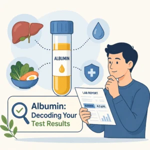
Alpha-1 antitrypsin is manufactured almost entirely in the liver. When liver function declines, production of this protein can fall along with production of albumin and other liver-made proteins, which is one reason clinicians rarely read a protein fraction in isolation. Understanding what an albumin blood level indicates alongside the alpha-1 band gives a much clearer signal than either value alone.
How alpha-1 globulins are measured on a lab report
The alpha-1 globulin figure comes from a serum protein electrophoresis, usually abbreviated SPEP on a requisition form. It is a standard blood draw from a vein in your arm, with no fasting requirement in most laboratories, although your clinician may ask you to fast if other tests are being drawn at the same time.
The laboratory reports the result in two ways. The absolute value gives the concentration in grams per deciliter, and the relative value gives the percentage of your total protein that sits in the alpha-1 band. Those two numbers can move in opposite directions. If your total protein drops sharply, the alpha-1 percentage can rise even though the absolute amount of alpha-1 protein has not changed at all. This is a common source of confusion when people compare two reports side by side.
Many reports also include a visual tracing, a curve with five peaks corresponding to the five bands. Clinicians look at the shape of that curve as well as the numbers, because an unusual spike or a flattened band can carry meaning that a single figure does not capture. If your report includes several unfamiliar values at once, our walkthrough of reference ranges, flags and trends on a lab report explains how to read them together.
Alpha-1 globulin normal range and how to read your result
For adults, the alpha-1 globulin normal range reported by the National Library of Medicine sits at roughly 0.2 to 0.4 grams per deciliter, equivalent to 2 to 4 grams per liter. Laboratories set their own ranges based on their equipment and the population they serve, so the range printed on your own report is the one that applies to you. Comparing a result against a different laboratory’s range is one of the most common ways people misread an otherwise unremarkable value.
The table below shows how the five bands on a protein electrophoresis relate to each other and what a shift in each one usually reflects.
| Band on the report | Main proteins it contains | What a shift often reflects |
|---|---|---|
| Albumin | Albumin | Nutrition, liver production, kidney protein loss, fluid balance |
| Alpha-1 | Alpha-1 antitrypsin, alpha-1 acid glycoprotein | Active inflammation when raised; an inherited antitrypsin deficiency when clearly low |
| Alpha-2 | Haptoglobin, ceruloplasmin, alpha-2 macroglobulin | Inflammation, red cell breakdown, kidney protein loss |
| Beta | Transferrin, complement proteins, some immunoglobulins | Iron status, lipid changes, some immune conditions |
| Gamma | Immunoglobulins (antibodies) | Infection, autoimmune activity, abnormal antibody-producing cells |
Because the bands are read together, a clinician comparing a raised alpha-1 result against the alpha-2 globulin fraction on the same report learns more than either number provides on its own. The same applies to the antibody band, and our guide covers what the gamma globulin fraction reveals when it sits outside its expected range.
The beta region deserves the same attention, and it splits in two on most reports: the beta-1 globulin fraction is dominated by transferrin, the protein that carries iron, so it tends to move with iron status rather than with inflammation.
Immediately beside it, the beta-2 globulin fraction carries complement proteins and part of the immunoglobulin load, so a shift there points in a different direction again.
What a high alpha-1 globulin level can mean
A raised alpha-1 globulin value is far more often a general signal of inflammation than a pointer to one specific illness. Both of the proteins in this band climb within roughly 24 to 48 hours of an inflammatory trigger, which means the value reflects what your body is doing right now rather than what it has been doing for months.
Common explanations include an active infection, a recent injury or surgery, a flare of an inflammatory condition such as rheumatoid arthritis or inflammatory bowel disease, pregnancy, and the use of estrogen-containing medications. Some cancers also raise the band, though a raised alpha-1 result on its own is a very weak cancer signal and is never used as a screening test for that purpose.
Clinicians almost always interpret a raised alpha-1 band alongside a dedicated inflammation marker. Checking a C-reactive protein blood result or reviewing an erythrocyte sedimentation rate measurement gives a much more direct read on inflammation than the protein fractions do, and those markers are cheaper and faster to repeat.
One practical point matters here. Because acute phase proteins fall as quickly as they rise, a raised alpha-1 value drawn during a chest infection is expected to normalize once the infection clears. Repeating the test several weeks after recovery, rather than immediately, usually answers the question of whether anything persistent is going on.
What a low alpha-1 globulin level can mean
A clearly low alpha-1 globulin result carries more specific weight than a raised one, because the band is small to begin with and there are only a few reasons for it to shrink.
The explanation clinicians look for first is alpha-1 antitrypsin deficiency, an inherited condition in which the liver produces a faulty version of the protein that struggles to leave liver cells and reach the bloodstream. The National Heart, Lung, and Blood Institute describes the consequence in two directions: the lungs lose their protection against enzymes released during inflammation, which can lead to emphysema in adulthood, and the misfolded protein accumulating inside liver cells can cause liver damage at any age. A protein electrophoresis showing a flattened alpha-1 band is often the first clue, and a direct alpha-1 antitrypsin level plus genetic testing confirms or rules out the diagnosis.
Other explanations for a low band include severe liver disease reducing overall protein production, significant protein loss through the kidneys or the gut, and malnutrition. Anyone investigating gut protein loss can review our guide to alpha-1 antitrypsin measured in a stool sample, a test used specifically to detect protein escaping through the intestinal wall.
Cleveland Clinic recommends testing for alpha-1 antitrypsin deficiency in anyone diagnosed with COPD, asthma or bronchiectasis, in anyone with unexplained liver disease, and in relatives of someone already diagnosed. That last point matters more than most people realize, since the condition is inherited and siblings share the highest risk. Our overview of chronic obstructive pulmonary disease and its symptoms explains why the lung link drives that recommendation.
When to talk to your doctor about an alpha-1 globulin result
Most alpha-1 globulin results that sit slightly outside the printed range need nothing beyond a repeat test at a sensible interval. A few situations genuinely warrant a conversation.
- Your alpha-1 band is clearly low rather than borderline, especially if you have unexplained breathlessness, a chronic cough, or abnormal liver enzymes.
- A close blood relative has been diagnosed with alpha-1 antitrypsin deficiency, whatever your own result looks like.
- You developed emphysema or COPD before age 50, or without a significant smoking history.
- Your alpha-1 value stays raised on a repeat test taken several weeks after any infection or injury has resolved.
- Several protein bands moved at once, particularly if the gamma band shows an unusual narrow spike.
- You have unexplained fatigue, weight loss, or bone pain alongside abnormal protein fractions.
Useful questions to bring to that appointment include which reference range your laboratory used, whether the result was affected by an illness you had around the draw date, and whether a direct alpha-1 antitrypsin measurement would answer the question more precisely than the electrophoresis band. If liver enzymes were flagged on the same panel, reading about alanine aminotransferase levels and what raises them helps you follow that part of the conversation.
How the alpha-1 band fits into a wider workup
A protein electrophoresis is rarely ordered to look at the alpha-1 band alone. Clinicians usually request it when total protein or the albumin to globulin ratio came back unexpected, when someone has unexplained bone pain or anemia, when nerve symptoms need investigating, or when a monoclonal protein needs ruling out.
That last purpose is worth understanding, because it explains why so many people encounter this test. When abnormal antibody-producing cells multiply, they release a single identical antibody that shows up as a sharp narrow spike, most often in the gamma region. That finding triggers further testing rather than a diagnosis on its own, and our guide to the kappa lambda light chain ratio covers the follow-up test used most often in that situation.
The practical takeaway is that the alpha-1 fraction is a supporting character in the report, not the lead. It adds context to the inflammation picture and it flags one specific inherited condition. It does not diagnose anything by itself, and a value a little outside the printed range in an otherwise well person is usually a reason to repeat the test rather than to escalate.
Latest scientific advances
Research over the past three years has sharpened two practical points about the proteins that make up the alpha-1 band.
The first concerns alpha-1 antitrypsin deficiency and how often it goes unrecognized. A review of the condition published in the Spanish journal Medicina Clínica set out the core problem plainly: the deficiency is regularly underdiagnosed, its severity varies a great deal from person to person, and treatment choices for the liver side of the condition remain limited (Dasí, 2023). The review also described the research directions being explored, including gene therapy and approaches that stop the faulty protein clumping inside liver cells, alongside the one approved drug treatment, augmentation therapy, which tops up the missing protein and can slow lung damage. What this means for you: if a lab report shows a low alpha-1 band and nobody has explained it, asking directly about a dedicated alpha-1 antitrypsin test is reasonable, because delayed recognition is the recognized weak point in how this condition is handled rather than a shortage of tests.
A second review, published in 2025, examined whether this deficiency is linked to asthma specifically. The authors found that published studies disagree widely with each other, that the differences are at least partly explained by inconsistent diagnostic criteria between research groups, and that current evidence does not establish the deficiency as a direct cause of asthma or show that augmentation therapy helps people with asthma who carry these gene variants (Lopez-Campos et al., 2025). They still supported testing people with severe or unusual asthma, in line with respiratory society guidance. What this means for you: carrying an alpha-1 gene variant does not mean your asthma was caused by it, but if your asthma is severe or behaves atypically, a one-time test is a sensible thing to ask about.
The third development concerns the other protein in the band. Alpha-1 acid glycoprotein has long been treated as a routine inflammation marker, and researchers have begun testing whether it tracks broader health outcomes. An analysis of a large United States national health survey found that among women aged 20 to 49, higher levels of this protein went together with a higher likelihood of frailty, meaning reduced physical resilience, and the marker distinguished frailty about as well as the more familiar high-sensitivity C-reactive protein test (Liu et al., 2025). This was a cross-sectional study, which means it captured a single snapshot in time and cannot show that one thing causes the other, and the authors said so themselves. What this means for you: this is early work in one population group, not a reason to request the test, but it supports the general principle that low-grade inflammation shows up across several blood proteins at once rather than in a single number.
Taken together, the recent literature reinforces a modest reading of this marker. The alpha-1 band is a useful pointer, particularly toward one inherited condition that benefits from early recognition, and it is not a standalone verdict on your health.
Glossary of key terms
| Term | Definition |
|---|---|
| Serum protein electrophoresis (SPEP) | A blood test that uses an electric current to separate blood proteins into five bands and measure how much protein sits in each one. |
| Globulin | One of the two main protein families in blood, alongside albumin, covering transport proteins, immune proteins and enzyme regulators. |
| Alpha-1 antitrypsin | A liver-made protein that protects lung tissue from enzymes released during inflammation, and the main component of the alpha-1 band. |
| Alpha-1 acid glycoprotein | Also called orosomucoid, a protein that rises during infection and inflammation and helps carry certain medications in the blood. |
| Acute phase reactant | A protein whose blood level rises or falls quickly in response to inflammation, usually within a day or two. |
| Reference range | The band of values seen in most healthy people for a given test at a given laboratory, printed alongside your result. |
| Monoclonal protein | A single identical antibody produced in excess by one clone of cells, seen as a narrow spike on an electrophoresis tracing. |
| Augmentation therapy | A treatment for alpha-1 antitrypsin deficiency that infuses the missing protein to help slow lung damage. |
| Emphysema | A form of lung damage in which the small air sacs are destroyed, reducing the surface available for oxygen exchange. |
FAQ
What causes a high alpha-1 globulin level?
The usual cause is active inflammation somewhere in the body. Infections, recent surgery or injury, flares of inflammatory conditions such as rheumatoid arthritis or inflammatory bowel disease, pregnancy and estrogen-containing medications all raise the band. Some cancers can raise it too, but the finding is far too nonspecific to be used as a cancer test. Because the proteins involved rise and fall quickly, a raised value often normalizes on its own once the underlying trigger resolves, which is why clinicians frequently repeat the test several weeks later rather than investigating immediately.
Does a high alpha-1 globulin result mean I have cancer?
On its own, no. A raised alpha-1 band is a general inflammation signal, and the overwhelming majority of raised results come from ordinary causes such as an infection or a recent injury. It is not a screening test for cancer and is never used as one. If your clinician is investigating cancer, the decision will rest on your symptoms, examination findings, imaging and targeted tests rather than on a protein fraction. A raised value with no symptoms and no other abnormal results is usually rechecked rather than escalated.
What does a low alpha-1 globulin level indicate?
A clearly low value most often points toward alpha-1 antitrypsin deficiency, an inherited condition in which the liver produces a faulty version of the protein that cannot reach the bloodstream properly. Other explanations include advanced liver disease reducing overall protein production, protein loss through the kidneys or the gut, and severe malnutrition. A low band is a reason to confirm with a direct alpha-1 antitrypsin measurement and, if that is low, genetic testing, rather than a diagnosis in itself.
Do I need to fast before a serum protein electrophoresis?
Most laboratories do not require fasting for this test, since the protein fractions are not meaningfully affected by a recent meal. However, this test is often drawn together with a metabolic or lipid panel that does require fasting, so follow whatever instruction your clinician or the laboratory gives you for the appointment as a whole. Tell the laboratory about any medications you take, particularly estrogen-containing ones, since some can shift protein levels.
Is the alpha-1 percentage or the absolute value more important?
Clinicians generally give more weight to the absolute value in grams per deciliter, because the percentage depends on your total protein. If total protein falls for an unrelated reason, the alpha-1 percentage can look raised even though the actual amount of alpha-1 protein has not moved. When you compare two reports over time, check whether you are looking at the same measure on both, and confirm that both came from the same laboratory using the same reference range.
Should my family be tested if I have alpha-1 antitrypsin deficiency?
This is worth discussing with your clinician. The condition is inherited, so siblings, children and parents carry a higher likelihood of having the same gene variants, and major clinical guidance supports offering testing to close relatives of someone diagnosed. Early identification matters because avoiding smoking and other lung irritants makes a substantial difference to long-term lung health, and monitoring can begin before symptoms appear. A genetic counselor can help family members decide what testing makes sense for them.
Sources
- MedlinePlus, National Library of Medicine — Protein electrophoresis – serum — MedlinePlus Medical Encyclopedia, reviewed 2024 — medlineplus.gov
- National Heart, Lung, and Blood Institute — Alpha-1 Antitrypsin Deficiency — NHLBI Health Topics, 2024 — nhlbi.nih.gov
- Cleveland Clinic — Alpha-1 Antitrypsin Deficiency: Symptoms and Treatment — Cleveland Clinic Health Library, 2024 — my.clevelandclinic.org
- Dasí F — Alpha-1 antitrypsin deficiency — Medicina Clínica, 2023 — doi.org/10.1016/j.medcli.2023.10.014
- Lopez-Campos JL, Muñoz-Sánchez B, Ferrer-Galván M, Quintana-Gallego E — Alpha-1 Antitrypsin Deficiency and Bronchial Asthma: Current Challenges — Biomolecules, 2025 — doi.org/10.3390/biom15060807
- Liu Y, Chen Y, Zhou L, et al. — Association between alpha-1-acid glycoprotein and frailty in US females aged 20-49 years: analysis of NHANES data (2015-2023) — Scientific Reports, 2025 — doi.org/10.1038/s41598-025-12188-z
Further reading
- Compare the neighboring band on the same electrophoresis report by reading our alpha-2 globulin results guide.
- Examine the antibody-carrying fraction of the report with our gamma globulin levels guide.
- Understand the protein figure that all five fractions are measured against in our total protein blood test guide.
- Check the marker clinicians rely on for a direct read on inflammation with our C-reactive protein results guide.
- Investigate protein leaking through the intestinal wall by reading our stool alpha-1 antitrypsin test guide.
Understand your lab results with BloodSense
Get your results interpreted in minutes
An alpha-1 globulin value means very little on its own, because it is one slice of a five-part protein picture that only makes sense when the bands are read together. Values such as total protein, albumin, the other globulin fractions, and a liver enzyme panel each add a piece of context that a single number cannot supply. BloodSense translates a full lab report into plain language, showing where each marker sits against its own reference range and how the results relate to one another. That makes it easier to arrive at your next appointment with specific questions rather than a page of unfamiliar figures.